Skin cancer (also known as “skin neoplasms”) are skin growths with differing causes and varying degrees of malignancy. The three most common malignant skin cancers are basal cell cancer, squamous cell cancer, and melanoma, each of which is named after the type of skin cell from which it arises. Skin cancer generally develops in the outermost layer of skin, so a tumour can usually be seen. This means that it is often possible to detect skin cancers at an early stage. Unlike many other cancers, only a small minority of those affected will actually die of the disease, though it can be disfiguring. Melanoma survival rates are poorer than for non-melanoma skin cancer, although when melanoma is diagnosed at an early stage, treatment is easier and more people survive.
Skin cancer is the most commonly diagnosed type of cancer. Melanoma and non-melanoma skin cancers combined are more common than lung, breast, colorectal, and prostate cancer.
Melanoma is less common than both basal cell carcinoma and squamous cell carcinoma, but it is the most serious. Most cases are caused by over-exposure to UV rays from the sun or sunbeds. Non-melanoma skin cancers are the most common skin cancers. The majority of these are basal cell carcinomas. These are usually localized growths caused by excessive cumulative exposure to the sun and do not tend to spread.
There are three main types of skin cancer: basal cell carcinoma (BCC), squamous cell carcinoma (SCC) and malignant melanoma.
Basal cell carcinomas are present on sun-exposed areas of the skin, especially the face. They rarely metastasize and rarely cause death. They are easily treated with surgery or radiation. Squamous cell carcinomas (SCC) are common, but much less common than basal cell cancers. They spread (metastasize) more frequently than BCCs. Even then, the rate of spread is quite low, with the exception of SCCs of the lip, ear, and in immunosuppressed patients. Melanomas are the least frequent of the 3 common skin cancers. They frequently metastasize, and could potentially cause death once they spread.
Less common skin cancers include: Dermatofibrosarcoma protuberans, Merkel cell carcinoma, Kaposi’s sarcoma, keratoacanthoma, spindle cell tumors, sebaceous carcinomas, microcystic adnexal carcinoma, Pagets’s disease of the breast, atypical fibroxanthoma, leimyosarcoma, and angiosarcoma.
The BCC and the SCC often carry a UV-signature mutation indicating that these cancers are caused by UV-B radiation via the direct DNA damage. However the malignant melanoma is predominantly caused by UV-A radiation via the indirect DNA damage.
Signs and symptoms
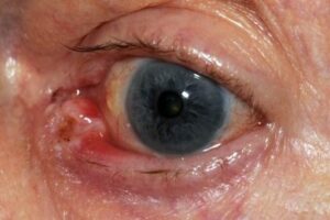
There are a variety of different skin cancer symptoms. These include changes in the skin that do not heal, ulcering in the skin, discolored skin, and changes in existing moles, such as jagged edges to the mole and enlargement of the mole.
If you are concerned about any lesion on your skin then an assessment by your health professional should be made.
Basal cell carcinoma usually presents as a raised, smooth, pearly bump on the sun-exposed skin of the head, neck or shoulders. Sometimes small blood vessels can be seen within the tumor. Crusting and bleeding in the center of the tumor frequently develops. It is often mistaken for a sore that does not heal. This form of skin cancer is the least deadly and with proper treatment can be completely eliminated.
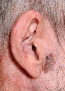
Most melanomas are brown to black looking lesions. Unfortunately, a few melanomas are pink, red or fleshy in color; these are called amelanotic melanomas. These tend to be more aggressive. Warning signs of malignant melanoma include change in the size, shape, color or elevation of a mole. Other signs are the appearance of a new mole during adulthood or new pain, itching, ulceration or bleeding. An often-used mnemonic is “ABCDE”, where A= asymmetrical, B= “borders” (irregular= “Coast of Maine sign”), C= “color” (variegated), D= “diameter” (larger than 6 mm—the size of a pencil eraser) and E= “evolving.”
Merkel cell carcinomas are most often rapidly growing, non-tender red, purple or skin colored bumps that are not painful or itchy. They may be mistaken for a cyst or other type of cancer.
Causes
Ultraviolet radiation from sun exposure is the primary cause of skin cancer. Other factors that play a role include:
Smoking tobacco
HPV infections increase the risk of squamous cell carcinoma.
Some genetic syndromes including congenital melanocytic nevi syndrome which is characterized by the presence of nevi (birthmarks or moles) of varying size which are either present at birth, or appear within 6 months of birth. Nevi larger than 20 mm (3/4″) in size are at higher risk for becoming cancerous.
Chronic non-healing wounds. These are called Marjolin’s ulcers based on their appearance, and can develop into squamous cell carcinoma.
Ionizing radiation, environmental carcinogens, artificial UV radiation (e.g. tanning beds), aging, and light skin color.
Sunscreen is effective and thus recommended to prevent melanoma and squamous cell carcinoma. Other advice to reduce rates of skin cancer includes: avoiding sunburning, wearing protective clothing, sunglasses and hats, and attempting to avoid periods of peak sun exposure.
The risk of developing skin cancer can be reduced through a number of measures including: Decreasing indoor tanning and mid day sun exposure and increasing the use of sunscreen
Avoiding the use of tobacco products
Treatment is dependent on type of cancer, location of the cancer, age of the patient, and whether the cancer is primary or a recurrence. Treatment is also determined by the specific type of cancer.
Treatment should be performed by a suitable trained health professional who can deal with all aspects of your skin cancer. Mr Stanley Loo is a plastic surgeon and is a specialist in treatment of all types of skin cancer and their reconstruction. Treatment of skin cancers are mostly surgery however other treatments may be beneficial in certain cases (Mr Loo will explain this to you in the consultation at East Care Medical Specialist Centre and perform the surgery at either Auckland City Surgical Services or Ormiston Hospital). Diagnosis is confirmed with an excisional biopsy (small surgical procedure). If the diagnosis of a skin cancer is confirmed further investigations may be required
Other treatment techniques include Mohs surgery, topical chemotherapy, immune response modifiers, cryotherapy (freezing the cancer), photodynamic therapy, and electrodesiccation and curettage.The goal of reconstructive surgery is restoration of normal appearance and function. Some of these techniques are indicated only for specific types of skin cancer. Mr Stanley Loo will discuss the best option for you in the consultation
The choice of technique in reconstruction is dictated by the size and location of the defect. Excision and reconstruction of facial skin cancers is generally more challenging due to presence of highly visible and functional anatomic structures in the face. Reconstruction is best performed by a specialist reconstructive plastic surgeon to give you the best result possible.
When skin defects are small in size, most can be repaired with simple repair where skin edges are approximated and closed with sutures. This will result in a linear scar. If the repair is made along a natural skin fold or wrinkle line, the scar will be hardly visible. Larger defects may require repair with a skin graft, local skin flap, pedicled skin flap, or a microvascular free flap. Skin grafts and local skin flaps are by far more common than the other listed choices.
Skin grafting is patching of a defect with skin that is removed from another site in the body. The skin graft is sutured to the edges of the defect, and a bolster is placed atop the graft for seven to ten days, to immobilize the graft as it heals in place. There are two forms of skin grafting: split thickness and full thickness. In a split thickness skin graft, a shaver is used to shave a layer of skin from the abdomen or thigh. The donor site, regenerates skin and heals over a period of two weeks. In a full thickness skin graft, a segment of skin is totally removed and the donor site needs to be sutured closed. Split thickness grafts can be used to repair larger defects, but the grafts are inferior in their cosmetic appearance. Full thickness skin grafts are more acceptable cosmetically. However, full thickness grafts can only be used for small or moderate sized defects.
Local skin flaps are a method of closing defects with tissue that closely matches the defect in color and quality. Skin from the periphery of the defect site is mobilized and repositioned to fill the deficit. Various forms of local flaps can be designed to minimize disruption to surrounding tissues and maximize cosmetic outcome of the reconstruction. Pedicled skin flaps are a method of transferring skin with an intact blood supply from a nearby region of the body.
In cases where there is spread to other areas, Mr Stanley Loo will discuss the treatment plan in the consultation.
Regular follow up is very important in the long term management of certain skin cancers and should be done by an appropriate health professional. Mr Loo will follow you up at his rooms (Auckland Plastic Surgery Group, Unit 6 / 31 Highbrook Drive, Highbrook, Auckland 2013) and arrange all appropriate investigation as required.
If you would like to discuss this further a consultation can be made with Mr Stanley Loo at Auckland Plastic Surgery Group, Unit 6 / 31 Highbrook Drive, Highbrook, Auckland 2013 Ph (09) 220 2580