A suspicious spot can create understandable uncertainty, particularly when a biopsy has confirmed skin cancer or your doctor has recommended removal. A skin cancer excision procedure is designed to remove the diagnosed lesion along with a planned margin of surrounding tissue, then send the specimen for laboratory assessment. The aim is complete treatment while preserving function, comfort and the best achievable scar.
Excision is a common and highly effective treatment for many skin cancers. However, the right approach depends on the type of cancer, its size and depth, its location, whether it has been treated before, and your individual health. A careful consultation gives you the opportunity to understand what is proposed and why.
Why excision may be recommended
Skin cancer excision is most commonly used for basal cell carcinoma, squamous cell carcinoma and melanoma. These cancers behave differently. Basal cell carcinomas usually grow locally and rarely spread elsewhere, but can cause significant local damage if left untreated. Squamous cell carcinomas have a greater potential to spread, particularly when they are large, deep, recurrent or located in higher-risk areas. Melanoma requires prompt, specialist-led assessment because its behaviour is closely related to the depth of the lesion.
In some circumstances, a biopsy has already established the diagnosis. In others, the entire lesion is removed because this is the most reliable way to diagnose and treat it. Non-surgical treatments may be suitable for selected superficial lesions, but excision has an important advantage: it provides tissue for pathology and allows the doctor to assess whether the cancer has been removed with clear margins.
The face, ears, scalp, hands, lower legs and areas close to the eyes, nose or lips need particular consideration. In these locations, surgical planning is about more than removing cancer. It must also protect important structures and achieve a wound closure that heals as well as possible.
Your consultation and surgical planning
Before surgery, your specialist will examine the lesion and review any biopsy or pathology report. They will discuss your medical history, medications, allergies, smoking status and previous scars or skin cancer treatment. Blood-thinning medication should never be stopped without advice from the clinician who prescribed it, but it may affect the timing and preparation for surgery.
The discussion should cover the expected scar, possible reconstruction, the type of anaesthetic, recovery, and the small but real risks of bleeding, infection, wound separation, altered sensation and an unfavourable scar. Most excisions are performed under local anaesthetic, meaning the area is numbed while you remain awake. Larger procedures, complex reconstructions or patient preference may mean sedation or a general anaesthetic is considered.
A margin is the rim of normal-looking skin removed around the visible lesion. It is not chosen arbitrarily. Recommended margins vary according to the diagnosis and risk features. A lesion with indistinct edges, a recurrent cancer or a melanoma may need a wider excision than a small, well-defined low-risk basal cell carcinoma. Your surgeon will balance oncological safety with the practical need to close or reconstruct the wound.
Planning the closure
A small wound can often be closed directly with stitches. If removing the lesion leaves too much tension, a skin graft or local flap may be recommended. A flap uses nearby skin and tissue, moved carefully while retaining its blood supply. It can be particularly useful where colour, thickness and contour matter, such as the nose, cheek or ear.
This is one reason a specialist assessment can be valuable. The method of closure is planned before the first incision, not treated as an afterthought once the cancer has been removed.
What happens during the skin cancer excision procedure
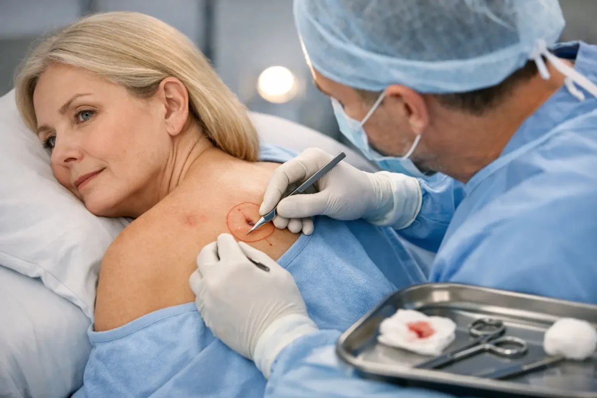
On the day, the lesion and planned margins are marked after confirming the site with you. This safety step is especially important if there are several lesions or if the area has become less visible after a biopsy.
For local anaesthetic surgery, you may feel a brief sting as the numbing medicine is injected. Once it has taken effect, you should feel pressure or movement but not sharp pain. Tell the surgical team immediately if you are uncomfortable. The skin is cleaned, sterile drapes are placed, and the lesion is removed with the intended margin of surrounding tissue.
The specimen is labelled and sent to a pathology laboratory. Depending on the wound and site, the surgeon may close the deeper layers with dissolving sutures and use fine stitches on the skin. A dressing is then applied. Straightforward procedures are often completed within an hour, although complex closures can take longer.
Occasionally, a planned direct closure is not appropriate once the true size of the defect is apparent. If this is a possibility, it should be discussed beforehand. Good surgical care includes having a clear plan while remaining prepared to make a safe adjustment if the findings require it.
Pathology results and clear margins
The laboratory examines the removed tissue under a microscope. The report confirms the diagnosis, describes relevant features of the cancer and comments on the excision margins. A clear margin means that no cancer is seen at the cut edge of the specimen. This is reassuring, although the meaning of a close margin can vary according to the cancer type and site.
If cancer cells extend to an edge, further treatment may be advised. This may involve a wider excision, a specialised margin-controlled technique, or occasionally radiotherapy or another treatment. It does not necessarily mean anything was done incorrectly. Some skin cancers have microscopic extensions beyond what can be seen on the surface, and pathology is the tool that identifies this.
Results are usually discussed at follow-up or communicated once they are available. Ask when and how you will receive your result before leaving the clinic. Waiting can be difficult, and knowing the next step often makes the period more manageable.
Looking after your wound
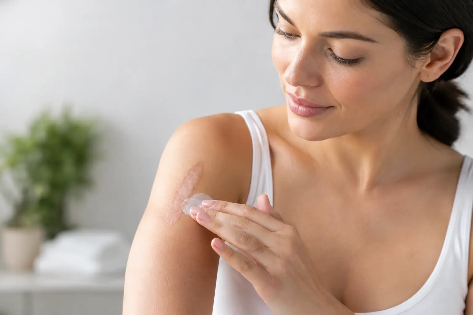
Your post-operative instructions will reflect the location and type of closure. Keep the dressing clean and dry for the period advised. After that, gentle washing may be appropriate, but avoid rubbing the wound. Do not apply creams, ointments or antiseptics unless your surgical team has recommended them, as some products can irritate healing skin.
Swelling, bruising and mild discomfort are common in the first few days. Elevating the area where practical, avoiding strenuous activity, and using the pain relief advised by your clinician can help. Procedures on the face often look more noticeable before they look better, and bruising may take one to two weeks to settle.
Contact your surgical team if you develop increasing rather than improving pain, spreading redness, warmth, pus-like discharge, fever, persistent bleeding or a dressing that becomes saturated. These symptoms do not always indicate infection, but they deserve prompt advice.
Stitches are removed according to the site and closure. Facial sutures are commonly removed sooner than stitches on the trunk or limbs, where healing is slower and tension can be greater. The scar will continue to mature for many months. Once the wound is fully healed, protecting it from sun exposure and following scar-care advice can support a better long-term result.
Follow-up is part of treatment
Having one skin cancer increases the chance of developing another. Follow-up therefore includes both checking the surgical site and helping you develop a practical surveillance plan. The frequency of review depends on the diagnosis, pathology findings, number of previous skin cancers and your personal risk factors.
Between appointments, become familiar with your skin. Seek assessment for a sore that does not heal, a spot that bleeds repeatedly, a changing mole, or a new lesion that continues to grow. Sun protection matters too: use shade, protective clothing, a broad-brimmed hat and suitable sunscreen as part of a broader routine, rather than relying on sunscreen alone.
The most reassuring treatment plan is one that gives equal attention to cancer clearance, careful reconstruction and the support you receive afterwards. If you have been advised to have an excision, a consultation with a specialist surgeon can help you understand your options and approach the procedure with clarity.