A new or changing mark on the skin can be easy to put off, particularly when it is small or painless. Yet a common and worthwhile question is: when should a skin lesion be removed? The answer depends on what the lesion looks and feels like, whether it is changing, where it sits on the body, and whether there is any concern that it could be cancerous. A careful specialist assessment can replace uncertainty with a clear plan.
When should a skin lesion be removed?
Removal may be recommended when a lesion has features that need diagnosis, causes persistent symptoms, is repeatedly injured, or is already known to be cancerous or pre-cancerous. Some lesions are removed for comfort or appearance, but medical safety always comes first.
Not every mole, skin tag, cyst or raised mark requires surgery. Many are harmless and can simply be monitored. However, a lesion should not be dismissed as benign without appropriate assessment simply because it has been present for a long time or does not hurt. Skin cancers can be subtle, and some lesions that appear routine need examination under magnification or laboratory testing to establish precisely what they are.
A lesion that is new or changing
Change is one of the most useful reasons to seek prompt review. This may include a mole that is getting larger, becoming raised, developing an irregular outline, or changing in colour. A lesion may also become darker, develop several shades, lose its usual pattern, or look noticeably different from the other marks on your skin.
The familiar ABCDE approach can help people notice potential melanoma warning signs: asymmetry, an irregular border, colour variation, a diameter that is increasing, and evolution or change over time. These features do not confirm melanoma, and melanomas do not always follow every rule. They do, however, justify an assessment.
New lesions in adulthood also deserve attention, especially if they continue to grow or look unusual. It is useful to take a clear photograph with the date noted, but photographs should support clinical review rather than replace it.
Bleeding, crusting, itching or failure to heal
A sore, spot or scaly patch that repeatedly bleeds, crusts, ulcerates, itches or fails to heal should be checked. Basal cell carcinoma and squamous cell carcinoma can sometimes resemble a persistent pimple, dry patch, wart or small cut. They may be painless, which is why waiting for discomfort is not a reliable approach.
A lesion that catches on clothing, jewellery, a razor or a comb may bleed because of friction alone. Even so, recurrent bleeding is worth discussing. A specialist can distinguish a straightforward traumatised benign lesion from a lesion that needs removal and histology.
A lesion that is painful, infected or repeatedly irritated
Benign lesions can still be troublesome. A skin tag at the neckline, a mole under a bra strap, or a cyst on the scalp may be regularly irritated during ordinary activities. Cysts can become inflamed or infected, causing tenderness, swelling and discharge. Removal may be considered once inflammation has settled, as operating through active infection can make healing more difficult and increase the chance of recurrence.
The trade-off is a scar. For a symptomatic lesion, a small, well-planned scar may be preferable to ongoing pain, bleeding or irritation. This decision should take account of the lesion’s location, your skin type, your healing history and the result you would consider acceptable.
Lesions that require urgent assessment
Most skin lesions do not require emergency treatment, but some should be assessed without delay. Arrange a prompt medical appointment if you notice a rapidly enlarging or very dark lesion, a mole changing over weeks or months, an unexplained non-healing sore, or a lesion that bleeds without being knocked.
Seek urgent care if an area becomes acutely red, hot, swollen and increasingly painful, particularly if there is fever or spreading redness. This may indicate an infection requiring treatment. If there is concern about a possible skin cancer, timely assessment matters, but it is also helpful to remember that an urgent review is not the same as a diagnosis. Many removed lesions prove to be benign.
Why removal can be the right diagnostic step
Sometimes a clinician can confidently identify a benign lesion from its appearance and recommend observation. In other cases, the only reliable way to reach a diagnosis is to remove all or part of the lesion and send the tissue to a laboratory for histological examination.
Histology examines the cells under a microscope. It can confirm whether a lesion is benign, pre-cancerous or cancerous, and, if cancer is present, provide information that guides further treatment. This is particularly valuable for pigmented lesions, changing moles and lesions with an uncertain diagnosis.
The method of removal depends on the clinical question. A shave excision may suit certain raised superficial benign lesions. A punch biopsy removes a small cylindrical sample and can be appropriate in selected cases. An excision biopsy removes the lesion with a margin of surrounding skin and is often used where complete assessment is needed. The most suitable technique should be chosen for both diagnostic accuracy and the best practical scar placement.
When removal is optional rather than medically necessary
A confirmed benign lesion may be removed for cosmetic reasons or because it causes inconvenience. Examples include stable moles in prominent areas, skin tags that catch on clothing, and harmless lumps that affect confidence. These are valid concerns, and they merit an honest discussion rather than being minimised.
However, elective removal should be approached with realistic expectations. Any procedure that cuts the skin leaves a scar, although careful planning and meticulous closure can help it settle well. Scars commonly look red or firm at first and may take many months to mature. Some areas, including the chest, shoulders and upper back, carry a higher risk of noticeable or raised scarring.
Before choosing cosmetic lesion removal, it is sensible to discuss the likely scar, the need for stitches, downtime, wound care and the possibility that a lesion could recur. If there is uncertainty about the diagnosis, tissue should usually be sent for histology rather than discarded.
What happens during a specialist lesion consultation?
A consultation begins with your history. You may be asked when you first noticed the lesion, whether it has changed, whether it bleeds or causes symptoms, and whether you have a personal or family history of skin cancer. Medication, smoking, sun exposure, prior scars and previous skin procedures can also affect planning.
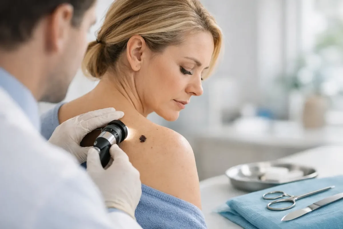
The lesion is then examined closely, often alongside the surrounding skin. You may be advised that removal is appropriate, that the lesion can be monitored, or that another type of assessment is needed first. A good consultation should make the reasoning clear, including what is known, what remains uncertain and what the scar may look like.
For many minor excisions, treatment is performed under local anaesthetic. The area is numbed, the lesion is removed, and the wound is closed where necessary. Most people can go home shortly afterwards. Depending on the site and closure, stitches may dissolve or need removal after a short period.
Recovery and aftercare matter
Good aftercare supports healing and helps reduce the risk of infection or poor scarring. You will receive individual instructions about keeping the wound clean and dry, dressings, exercise, showering and when to return for review. It is usually wise to avoid stretching or strenuous activity that pulls on the wound while it is healing.
Contact your surgical team if you develop increasing pain, spreading redness, pus-like discharge, significant swelling, wound separation or a fever. Once the wound has healed, protecting the scar from sun exposure can reduce prolonged redness and pigment change.
If histology is performed, the result should be explained in plain language. Where further treatment or surveillance is needed, this should be arranged with clear guidance rather than leaving you to interpret a laboratory result alone.
Do not wait for certainty
You do not need to decide for yourself whether a mark is harmless before arranging an assessment. The sensible threshold is lower: if a lesion is changing, bleeding, persistent, symptomatic or simply worrying you, have it examined. A calm, informed review can determine whether observation is safe or whether removal offers the reassurance, comfort or treatment you need.