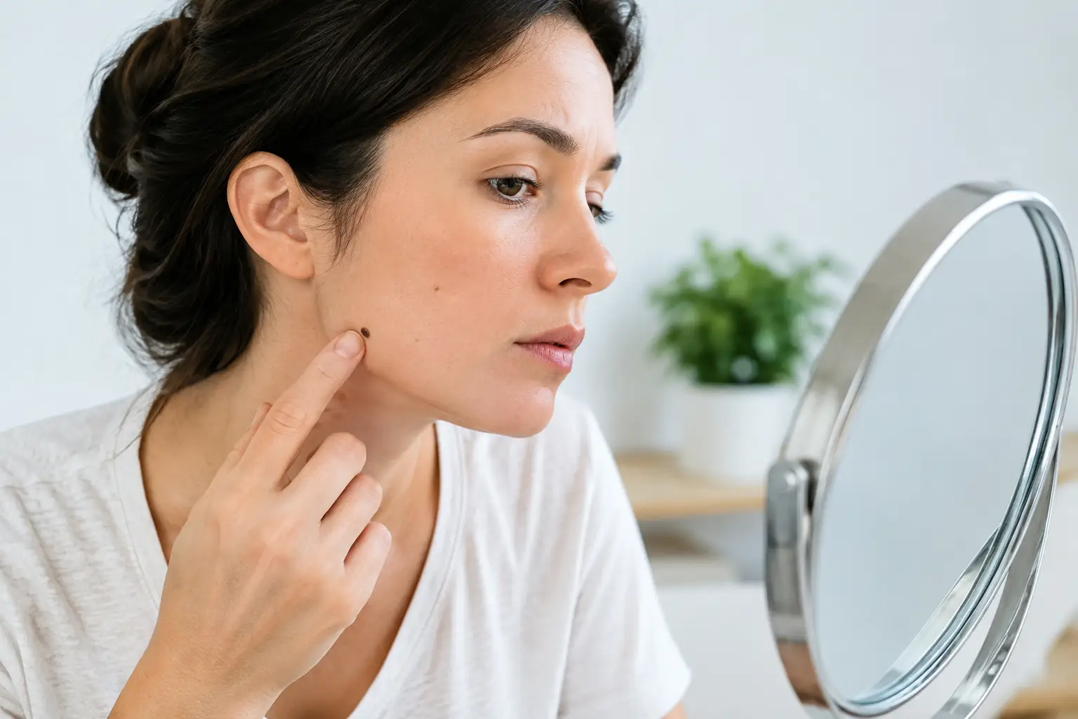
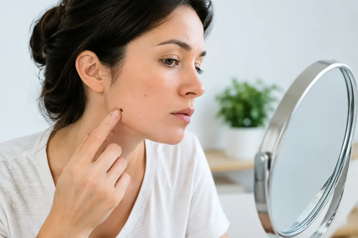
A mole does not need to be painful, bleeding or dramatically raised to deserve attention. Often, the earliest concern is simply that it looks different from the other marks on your skin, or that it has changed in a way you cannot explain. Knowing how to assess suspicious moles can help you act promptly, while recognising that a home check is not a diagnosis.
Most moles are harmless. They may be flat or raised, light brown, dark brown or close to skin colour, and many remain unchanged for years. The purpose of checking your skin is not to create alarm over every mark. It is to notice new, evolving or unusual lesions and arrange an expert assessment when one is needed.
How to assess suspicious moles at home
Choose good natural light and use a full-length mirror alongside a hand mirror for difficult areas. Check your face, scalp, neck, torso, arms, hands, legs and feet. Do not overlook the soles of your feet, between the toes, under the nails, or the genital area. If possible, ask a partner, friend or family member to inspect your back and scalp.
A monthly check is reasonable for many adults, particularly if you have numerous moles, fair skin, a history of significant sun exposure, or a close relative with melanoma. For people at higher risk, a clinician may recommend a more individual schedule.
When looking at a particular mole, use the ABCDE approach as a practical prompt:
- A – Asymmetry: One half of the mole does not match the other in shape, thickness or colour.
- B – Border: The edge is irregular, blurred, scalloped or poorly defined rather than smooth and even.
- C – Colour: There is more than one shade, such as brown, black, red, white, blue or grey, or the colour has changed.
- D – Diameter: A lesion wider than about 6 mm can warrant review, although smaller melanomas can occur.
- E – Evolving: The mole is changing in size, shape, colour, surface or sensation. This is often the most useful warning sign.
The ABCDE rule is a helpful screening tool, not a reason to wait until every feature is present. A lesion with one concerning characteristic, particularly clear change over weeks or months, should be assessed.
Look for the “ugly duckling”
Your skin usually has a pattern. You may have many freckles or moles, but most will tend to resemble one another in colour and appearance. An “ugly duckling” is a mark that looks noticeably different from the rest.
It may be darker, pinker, larger, more raised or more irregular than your usual moles. It may also be a new lesion that appears after adulthood and does not fit your normal skin pattern. This comparison can be especially useful for people with many moles, where applying the ABCDE rule to every individual mark can feel difficult.
Not every unusual-looking lesion is cancerous. Seborrhoeic keratoses, sunspots, harmless raised moles and benign vascular lesions can all look unfamiliar. However, it is safer to have a clinician examine a changing or distinctive lesion than to make assumptions based on appearance alone.
Changes that need prompt medical advice
Arrange an appointment with your GP, dermatologist or appropriate skin specialist if a mole is new and changing, or if an existing mole has altered. Changes in colour, outline, height or size matter, but so do symptoms. Itching, tenderness, crusting, repeated bleeding, ulceration or a sore that does not heal should be checked.
Some melanomas do not follow the classic image of a dark, irregular flat mole. Nodular melanoma, for example, may develop as a rapidly growing dome-shaped lump that is firm, raised and may be black, brown, red, pink or skin coloured. Speed of change is particularly significant.
Seek more urgent advice if a lesion is bleeding without a clear injury, growing quickly, becoming persistently painful, or developing a dark streak beneath a nail. Do not try to remove, cut, freeze or treat a suspicious mole yourself. Home treatments can cause inflammation and scarring, and may delay a reliable diagnosis.
Photographs can help, but have limits
A clear photograph can be useful for recording a lesion and noticing whether it is changing. Take the image in consistent lighting, include a ruler or coin for scale, and record the date. Repeat photographs can give your clinician helpful context, particularly if the mole is in a difficult-to-see area.
However, photographs cannot show all the details a specialist needs to assess. Lighting can distort colour, camera images cannot assess texture accurately, and an image cannot replace examination with dermoscopy. Dermoscopy is a close assessment using a magnifying device and specialised light, allowing the clinician to evaluate structures beneath the skin surface that cannot be seen with the naked eye.
Avoid relying on mobile applications to decide that a mole is safe. Some tools may encourage skin awareness, but they cannot provide the certainty of a clinical examination or, where appropriate, laboratory analysis.
Your personal risk affects the threshold for review
Anyone can develop skin cancer, including people with darker skin tones and those who do not burn easily. That said, earlier review is sensible if you have previously had melanoma or another skin cancer, have a first-degree relative with melanoma, have a large number of moles, or have a history of intense intermittent sun exposure and sunburn.
Immunosuppressive medicines, previous radiotherapy and a history of using sunbeds may also increase risk. If you are uncertain whether your medical history changes your risk, raise it during your appointment. A clinician can advise whether routine skin surveillance would be appropriate for you.
What happens at a specialist skin assessment?
A consultation begins with a careful history. You may be asked when you first noticed the lesion, whether it has changed, whether it causes symptoms, and whether there is relevant personal or family history. The clinician will examine the lesion, and may examine other areas of skin to identify additional lesions requiring attention.
If a mole appears benign, reassurance and observation may be all that is required. If it is uncertain or suspicious, removal or biopsy may be advised. The recommendation depends on the lesion, its location, your medical history and the clinical findings.
Where excision is recommended, the lesion is usually removed under local anaesthetic and sent for histological examination. This means a pathologist examines the tissue under a microscope to establish the diagnosis. The final pathology result, rather than appearance alone, guides any further treatment. Your surgeon should explain the expected scar, wound care, recovery and possible risks before proceeding, so you can make an informed decision.
Protecting your skin while remaining alert
Regular skin checks work best alongside sensible sun protection. Use broad-spectrum sunscreen, seek shade during the strongest sun, wear protective clothing and avoid sunbeds. Sun protection reduces future ultraviolet damage, but it does not remove the need to assess a changing lesion.
Try not to become so vigilant that every normal freckle becomes a source of worry. The practical question is whether a mark is new, changing, symptomatic or clearly unlike your other moles. If the answer is yes, a professional assessment offers clarity and, where treatment is needed, the best opportunity for early care.
If a mole has caught your attention more than once, it is worth arranging an appointment. Reassurance is valuable when a lesion is harmless, and timely assessment is the sensible next step when it is not.