A mole that repeatedly catches on clothing, bleeds after minor friction, changes in appearance or simply makes you self-conscious deserves proper attention. Mole removal options should always begin with a clinical assessment, rather than choosing a treatment based on appearance alone. The priority is to establish whether a lesion is likely to be benign and whether removal is medically appropriate, cosmetically worthwhile or both.
For many patients, removal is a straightforward minor procedure performed under local anaesthetic. However, the best technique depends on the mole’s size, depth, location, colour, shape and clinical features, as well as your preference regarding scars, recovery and certainty of diagnosis.
Why assessment comes before mole removal
Most moles are harmless. Some have been present for years without causing concern, while others become irritating because they rub against a bra strap, collar, waistband or shaving area. A specialist examination helps distinguish these common benign lesions from marks that need further investigation.
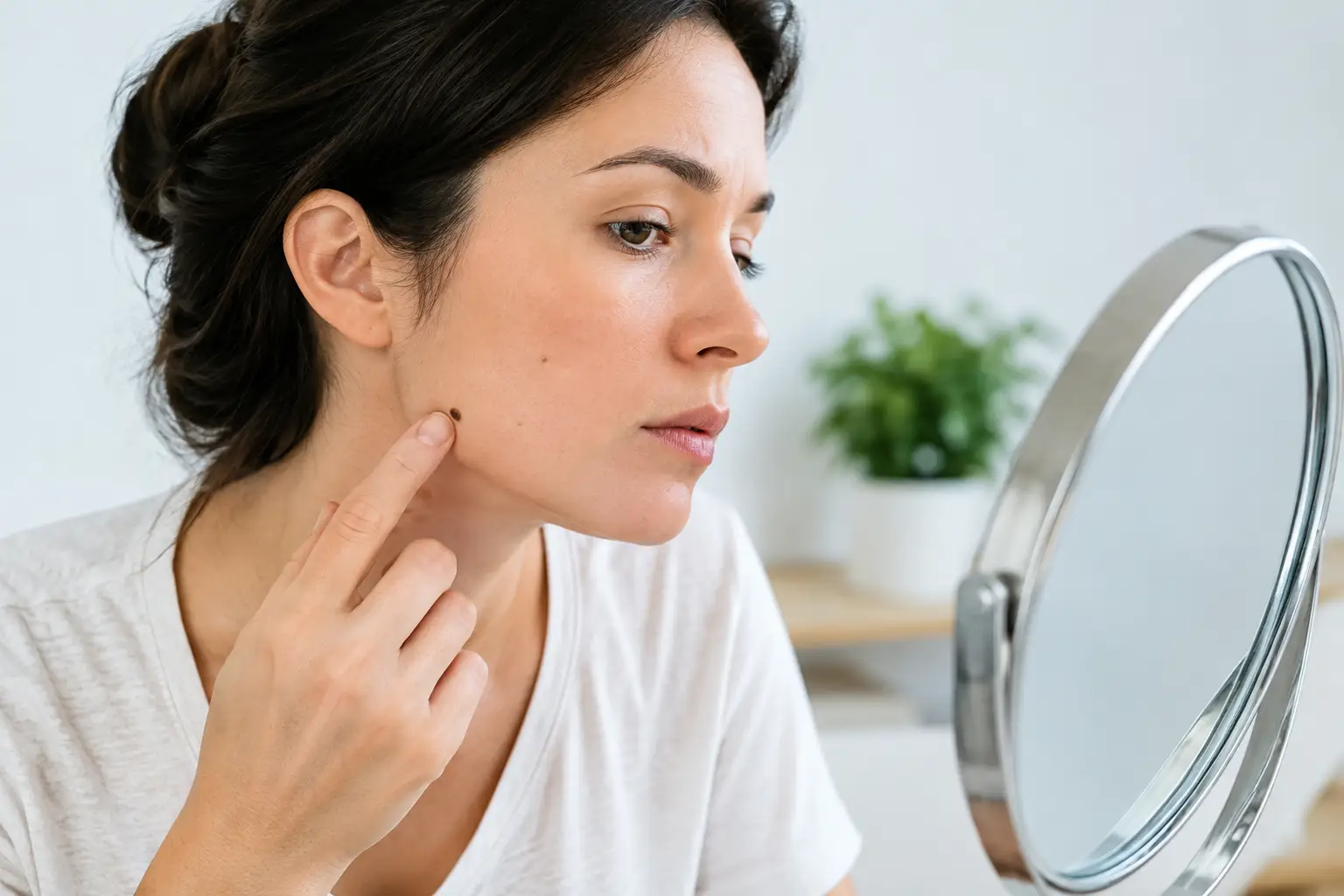
It is sensible to arrange a prompt medical review if a mole is new or changing, particularly if it is becoming larger, asymmetric, irregular at the edge, varied in colour, itchy, painful, crusted or bleeding without being knocked. A lesion that looks noticeably different from your other moles also warrants assessment. These features do not automatically mean skin cancer, but they should not be ignored.
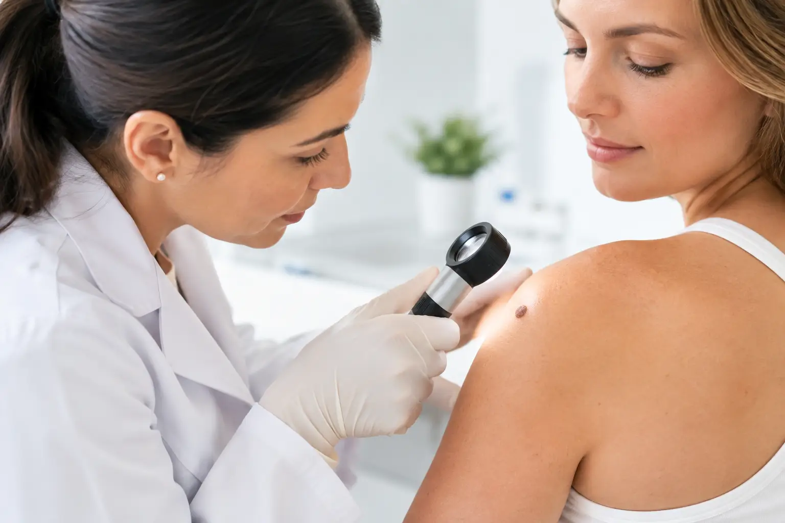
During a consultation, the clinician will ask when you first noticed the mole, whether it has altered, whether there is a personal or family history of skin cancer, and how the lesion affects you. Examination may include dermoscopy, a close assessment using magnification and specialised lighting. If removal is advised, the reasons for the chosen approach should be explained clearly before treatment proceeds.
Mole removal options and when they may be used
Monitoring rather than removal
Not every benign mole needs to be removed. If a lesion is clinically reassuring, causes no symptoms and is not troubling you cosmetically, monitoring may be the most appropriate option. This avoids an unnecessary procedure and the permanent scar that any skin removal can leave.
Monitoring may involve photographs, measurements and guidance on changes to watch for. It is particularly useful when the diagnosis is clear but the mole is in an area where scarring could be more noticeable, such as the face, chest or shoulder.
Shave excision
Shave excision is often suitable for a raised mole that appears benign and sits mainly above the skin’s surface. After local anaesthetic is given, the lesion is carefully removed level with, or just below, the surrounding skin. Stitches are not usually required.
The wound generally heals over one to two weeks, depending on its size and location. This technique can offer a good cosmetic result for selected raised lesions, particularly where a linear scar from stitches may not be preferable. The trade-off is that deeper mole cells can occasionally remain, meaning the mole may recur. It may also leave a slightly lighter, darker or flatter area of skin.
Where there is any diagnostic uncertainty, the removed tissue can be sent for laboratory examination. A specialist will advise whether shave excision is appropriate or whether full-thickness removal is safer.
Surgical excision
Surgical excision removes the entire mole, including its deeper component, with a small margin of surrounding skin where needed. The wound is then closed with stitches, usually creating a fine linear scar. It is commonly recommended for flat or deeper moles, lesions with concerning features, or moles where complete removal and histological analysis are important.
Although the scar is longer than the original mole, careful planning follows natural skin lines where possible. The precise shape and length depend on the lesion’s position and how much skin can be closed without tension. Stitches may be dissolvable or removed at a follow-up appointment, often after around one to two weeks depending on the body area.
Excision offers the clearest answer when a diagnosis needs confirmation because the laboratory can assess the whole lesion. If abnormal cells are identified, the result helps guide whether any further treatment is required.
Punch excision
For some small, deep lesions, a punch excision may be appropriate. This uses a circular instrument to remove the mole through the full thickness of the skin. The resulting wound may be closed with one or two stitches.
This can be an efficient option for selected small moles, but it is not ideal for every location or lesion shape. As with other procedures, the aim is to balance complete removal, diagnostic confidence and the likely appearance of the scar.
Why laser or freezing may not be the right answer
Patients sometimes ask about laser treatment, cautery or freezing because these methods can sound quick and scar-free. They are not automatically suitable for pigmented lesions or moles. If a mole has not been properly assessed, destroying it without obtaining tissue for analysis can make diagnosis more difficult.
A procedure that sounds less invasive is not necessarily the safest or most cosmetic choice. For a lesion that is raised and clearly benign, a minor surgical approach may offer a more predictable result. For a suspicious lesion, excision and laboratory analysis are generally more appropriate than treatments that destroy tissue.
What to expect on the day
Mole removal is usually carried out as an outpatient procedure under local anaesthetic. You remain awake, but the area is numbed before treatment begins. You may feel the initial sting of the anaesthetic and some pressure during the procedure, but you should not feel sharp pain.
The skin is cleaned, the planned removal is marked and the mole is removed using the agreed technique. If stitches are required, a dressing is applied and you will receive advice on caring for the wound. Most people can return home shortly afterwards and resume light daily activities, although strenuous exercise, swimming and friction on the area may need to be avoided initially.
If the lesion is sent for histology, you should be told how and when results will be communicated. It is reasonable to ask whether the mole will be analysed, what the likely scar will look like, when stitches need removing and who to contact if you have concerns during healing.
Healing, scars and aftercare
Every skin procedure leaves some form of mark. The goal is not to promise a scar-free result, but to choose an approach that provides an appropriate clinical outcome with the best achievable scar for your skin and the treatment area.
In the first few weeks, a scar may look pink, firm or slightly raised. It usually settles gradually over months, although scar maturation can take a year or longer. Areas under movement or tension, such as the shoulders, chest and upper back, can be more prone to wider or more noticeable scars. Your individual healing history also matters.
Keeping the wound clean and dry as advised, avoiding picking and protecting the healed scar from sun exposure are simple but valuable steps. Once the wound has fully healed, scar care may include massage or silicone-based products if recommended. Contact the clinic if you develop increasing redness, warmth, swelling, discharge, worsening pain or a fever, as these can indicate infection.
Choosing the right practitioner
For a lesion that is changing, symptomatic or diagnostically uncertain, clinical judgement matters as much as the removal itself. A consultation with a suitably qualified doctor or specialist allows the lesion to be assessed properly, the removal method to be tailored to you and tissue to be examined where appropriate.
At Stanley Loo’s practice, the emphasis is on consultant-led assessment, clear explanation and attentive follow-up. You should feel able to discuss both the medical and cosmetic aspects of treatment, including the possibility of a scar, recurrence, laboratory testing and the expected recovery.
The right choice is rarely the quickest advertised treatment. It is the option that gives you confidence that the mole has been assessed carefully, removed appropriately and supported with clear aftercare.