A mole that repeatedly catches on clothing, a lump that has started bleeding, or a mark that simply does not look like the rest of your skin deserves proper assessment. The best options for skin lesion removal are not the same for every lesion. The right approach depends on what the lesion is, where it sits, whether it is changing, and whether laboratory analysis is needed.
For many patients, the concern is practical or cosmetic: irritation from shaving, discomfort from spectacles, or a visible lesion affecting confidence. For others, removal is recommended because a lesion may be suspicious for skin cancer. A consultation with an appropriately qualified clinician provides the opportunity to assess the skin carefully, discuss the likely diagnosis and explain the treatment, scar and recovery considerations before a decision is made.
When a skin lesion should be assessed
Not every mole, skin tag or raised mark needs to be removed. Many harmless lesions can be safely monitored. However, a new lesion or a change in an existing one should be checked rather than treated at home or removed purely for cosmetic reasons without a diagnosis.
Arrange a medical assessment promptly if a lesion has changed in size, shape or colour; has an irregular border; develops several different colours; bleeds, crusts or ulcerates without an obvious reason; becomes persistently itchy, painful or tender; or does not heal. These changes do not always mean skin cancer, but they warrant clinical review.
A specialist will consider your medical history, sun exposure, previous skin cancers and family history alongside the appearance of the lesion. In some circumstances, dermoscopy, a close examination using a magnifying light, can provide further detail. If there is concern, the removed tissue may be sent for histological examination, where a pathologist examines it under a microscope.
Best options for skin lesion removal: treatment choices
The most suitable technique is guided by the diagnosis. It is also influenced by the lesion’s size, depth and location, as well as the need to obtain a complete tissue specimen. A treatment that is appropriate for a soft skin tag may not be appropriate for a changing pigmented mole.
Monitoring rather than removal
Observation can be the best option when a lesion appears benign, is not causing symptoms and is unlikely to cause future problems. Your clinician may advise taking photographs or monitoring for particular changes. This avoids a procedure and the resulting scar.
Monitoring is not a substitute for review if the lesion changes. If you are worried, or cannot confidently keep track of a lesion in a difficult-to-see area such as the back or scalp, it is sensible to seek reassessment.
Shave excision
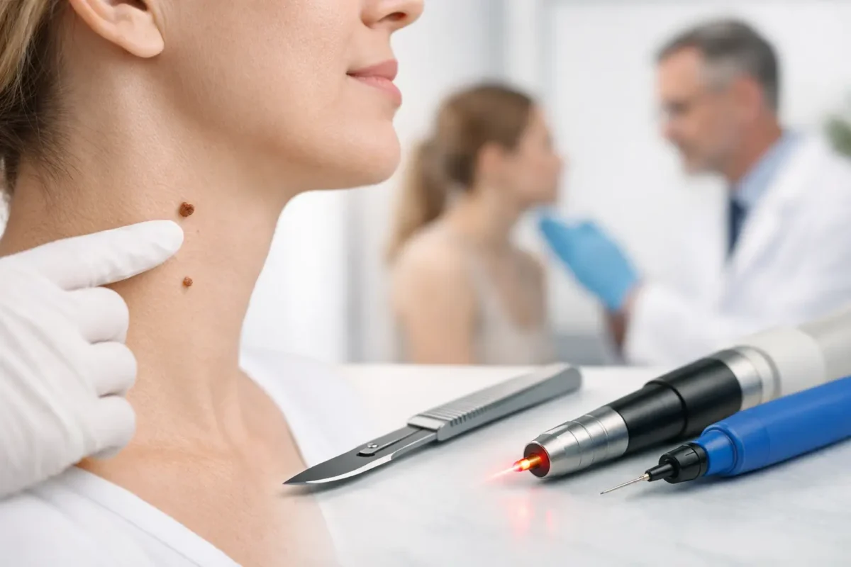
A shave excision removes a raised lesion at or slightly below the level of the surrounding skin. It is commonly used for selected benign moles, skin tags and some other superficial growths. Local anaesthetic is used, and stitches are often not required.
This can offer a straightforward recovery, although the area will need wound care while it heals. A flat, paler or slightly indented mark may remain. Shave excision is not suitable where a lesion is suspicious, deep, or where full-thickness assessment and clear margins are required.
Formal surgical excision
Surgical excision involves removing the lesion completely, usually with a narrow margin of normal-looking skin where clinically appropriate. The wound is then closed with stitches. This is often the preferred approach for suspicious pigmented lesions, lesions requiring histology, skin cancers and deeper lumps.
The key advantage is that it provides an intact specimen for laboratory assessment and can allow the clinician to assess whether a lesion has been fully removed. The trade-off is a linear scar and a slightly longer period of care while stitches remain in place. Thoughtful planning of the incision can help place the scar along natural skin lines where possible, but no procedure can guarantee an invisible scar.
Curettage, cautery or cryotherapy
Some superficial benign lesions can be treated by curettage, which gently removes tissue, sometimes combined with cautery to control bleeding. Cryotherapy uses controlled freezing and may be used for selected lesions such as certain warts or sun-related surface changes.
These techniques can be useful in the right circumstances, but they do not always produce a specimen suitable for histology. They are therefore generally avoided when the diagnosis is uncertain or skin cancer is suspected. Healing may involve a scab, temporary colour change or, occasionally, a small scar.
Laser treatment
Laser treatment may be considered for carefully selected benign surface lesions or areas of pigmentation. It is not automatically the most precise or scar-free option, despite its popularity in cosmetic advertising. Some lesions can recur after laser treatment, and laser may destroy tissue that would otherwise have been examined under a microscope.
For a changing mole, an undiagnosed pigmented lesion or a lesion with features concerning for skin cancer, laser treatment is usually not the appropriate first step. Establishing a diagnosis comes before choosing a cosmetic technique.
What determines the right procedure
The consultation should be more than a quick decision to remove a mark. A careful plan considers whether the lesion needs testing, the best way to minimise unnecessary scarring, and the effect of the procedure on nearby structures. This matters particularly around the eyelids, lips, nose, ears, hands and joints, where function and wound tension must be respected.
Your clinician should explain whether the goal is diagnosis, complete treatment, symptom relief, cosmetic improvement or a combination of these. For example, a benign lesion on the face may be removed in a way that prioritises scar placement, while a suspected melanoma requires an approach led first by accurate diagnosis and appropriate excision.
It is also worth discussing medication, allergies, smoking or vaping, previous problems with scars, blood-thinning medicines and any history of poor wound healing. These factors can affect procedural planning and aftercare.
What to expect from lesion excision and recovery
Most minor lesion removals are performed under local anaesthetic. You will be awake but the area should be numb during the procedure. Some pressure or movement may be noticeable, but sharp pain should not be expected. The exact appointment length depends on the site and complexity of the lesion.
Afterwards, a dressing may be applied and you will receive specific instructions about keeping the wound clean, showering, activity and when stitches should be removed. Mild discomfort, bruising or swelling is common in the early days and is usually manageable with simple pain relief, if suitable for you.
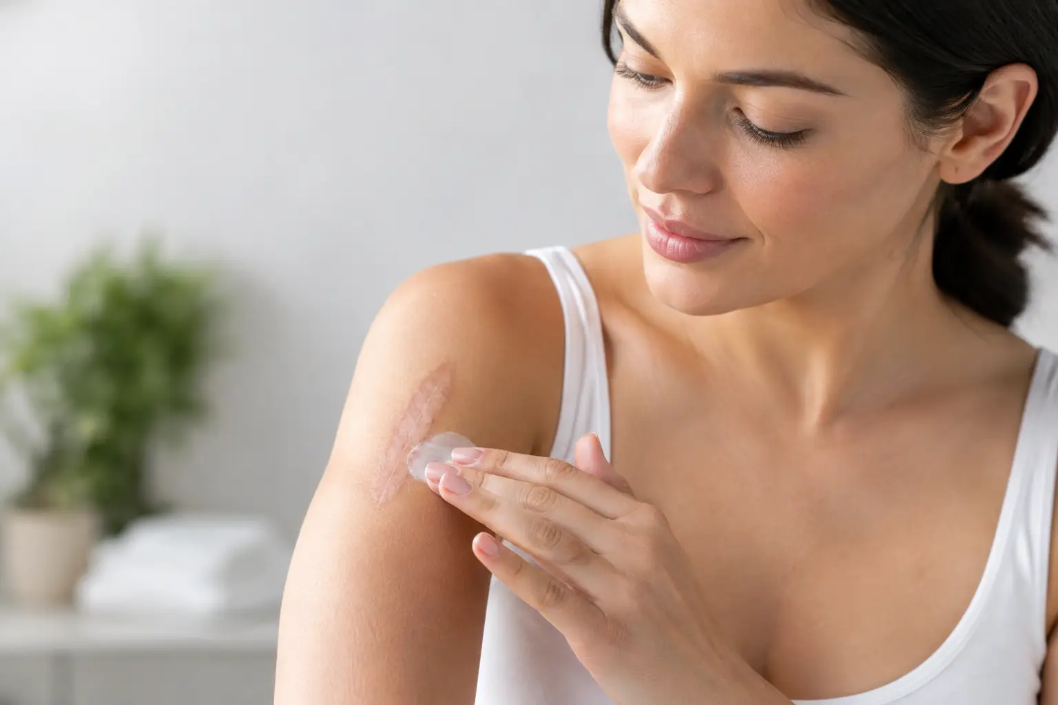
Scar maturation takes time. A new scar often looks pink or firm before gradually settling over several months. Protecting the area from sun exposure, avoiding unnecessary tension on the wound and following the recommended care plan can support healing. If pathology is being performed, the result should be discussed with you and any further treatment explained clearly.
Seek advice if you develop increasing redness, warmth, swelling, discharge, worsening pain, fever, wound separation or persistent bleeding. Early review helps address concerns before they become more significant.
Choosing care with confidence
The safest choice is not necessarily the quickest or least expensive treatment advertised for a skin concern. A proper assessment helps ensure that a lesion is not treated in a way that delays diagnosis or compromises the ability to examine it histologically.
At Stanley Loo, lesion removal is planned around the individual patient, with clear discussion of the diagnosis, procedure, scar expectations and post-operative care. If a lesion is changing, uncomfortable or troubling you, arranging a consultation can replace uncertainty with a considered plan for your skin and your peace of mind.