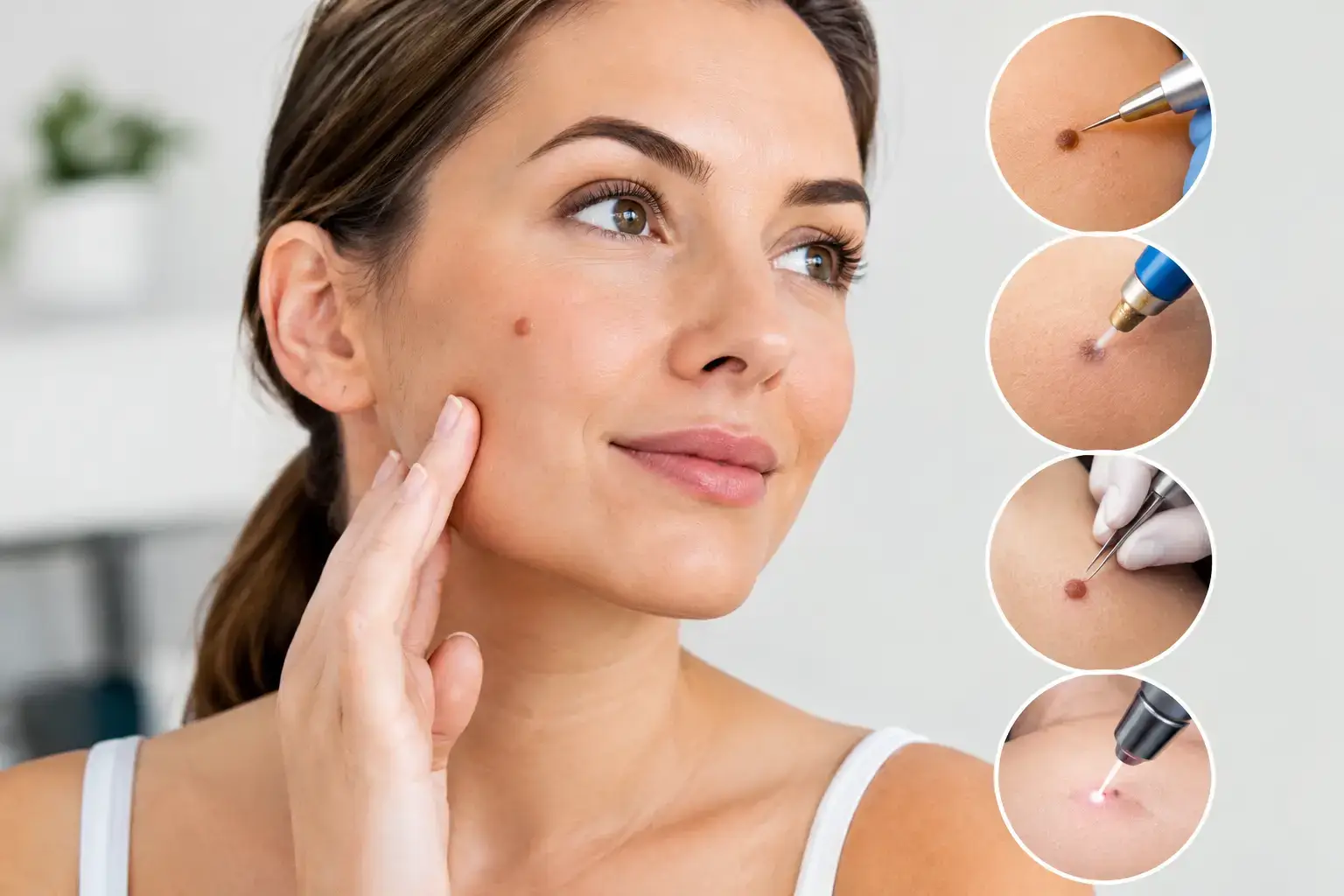
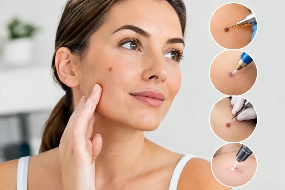
A mole may be completely harmless, yet still cause concern because it catches on clothing, is repeatedly irritated when shaving, affects confidence, or has changed in appearance. The top mole removal methods are not interchangeable. The right approach depends first on whether the lesion needs diagnostic assessment, then on its size, depth, location and the result you hope to achieve.
For any new, changing, bleeding or unusually coloured mole, removal should never be treated as a purely cosmetic decision. A careful clinical examination is the starting point. Where there is any concern, the lesion may need to be removed in a way that allows it to be examined under a microscope.
Why an assessment comes before mole removal
Moles are common collections of pigment-producing cells. Most are benign, but some skin lesions can resemble one another, and occasional changes may indicate skin cancer. A specialist consultation provides the opportunity to discuss when you first noticed the lesion, whether it has altered, and whether there is a personal or family history of skin cancer.
The examination considers features such as asymmetry, irregular borders, variation in colour, increasing diameter and evolution over time. These features are useful prompts for assessment, not a diagnosis that can be made from a photograph alone. Your clinician may recommend monitoring, removal, or referral for further assessment depending on the findings.
This discussion also matters for the cosmetic outcome. Removing a mole replaces it with a scar. In experienced hands, scar placement and wound closure are planned carefully, but no procedure can guarantee an invisible mark. Skin type, body site, healing tendency and aftercare all influence the final result.
Top mole removal methods explained
Surgical excision
Surgical excision is often the preferred option when a mole is raised, deep, clinically atypical, or needs histological analysis. The area is numbed with local anaesthetic. The surgeon removes the entire lesion, usually with a small margin of normal-looking skin where appropriate, and closes the wound with sutures.
The specimen can then be sent to a laboratory for histology. This is a significant advantage when there is any uncertainty about the diagnosis. Excision also offers the best chance of complete removal for many moles, particularly those that extend below the surface of the skin.
The trade-off is a linear scar and, depending on the site, a short period of activity modification while the wound heals. Sutures may be dissolvable or may need removal at a follow-up appointment. For a suspicious lesion, the priority is complete, appropriate removal and an accurate diagnosis rather than minimising the initial scar at all costs.
Shave excision
A shave excision may be suitable for selected benign-looking moles that project above the skin. After local anaesthetic, the visible raised portion is carefully removed level with, or just below, the skin surface. Stitches are often not required, and the area heals with a dressing.
This can be a practical option for certain raised moles on the face, scalp or body, particularly where a straight-line scar may be less desirable. Healing is usually straightforward, although the site may remain pink for several weeks and can leave a lighter, darker or slightly indented mark.
A shave technique is not appropriate for every mole. It may not remove cells located deeper in the skin, meaning pigment can recur over time. If histology is required or the lesion has concerning features, full excision may be recommended instead.
Punch excision
Punch excision uses a small circular instrument to remove a discrete lesion through the full thickness of the skin. It is most useful for small moles where a neat circular removal is appropriate. The wound may be closed with one or two stitches, or occasionally allowed to heal naturally depending on its size and location.
As with standard excision, the tissue can be sent for histology. The resulting scar is usually small, but it can widen or become more noticeable in high-tension areas such as the shoulders, chest or upper back.
Observation and monitoring
Not every mole should be removed. If a lesion appears benign and is not causing physical or emotional discomfort, monitoring may be the most sensible course. This avoids an unnecessary procedure and a permanent scar.
Monitoring does not mean ignoring a mole. Take note of changes in size, shape, colour, symptoms such as itching or bleeding, and the appearance of a new lesion that looks different from your other moles. Seek medical advice promptly if you notice a change. Your clinician can advise whether clinical photographs or review at a later date would be useful.
Methods that are not usually appropriate for moles
Laser treatment, cautery and cryotherapy are used for particular skin concerns, but they are not generally first-line treatments for a pigmented mole that requires diagnosis. These methods can destroy tissue, leaving no specimen for histology. They may also lead to incomplete treatment or pigment recurrence.
Over-the-counter mole-removal products should be avoided. They can cause burns, infection, scarring and delayed assessment of a potentially serious lesion. A lesion should not be self-treated simply because it appears small or familiar.
Choosing between mole removal options
The best method is guided by the lesion rather than a one-size-fits-all preference. A flat or changing pigmented lesion may need formal excision and histology. A small, clearly benign raised mole may be suitable for shave removal. A small, well-defined lesion may be considered for punch excision.
Location is equally relevant. The face, eyelids, scalp, chest, back and limbs heal differently and are exposed to different forces. A surgeon will consider natural skin creases, tension lines and the visibility of the scar. Previous keloid or hypertrophic scarring should be discussed, as should medications that affect bleeding or healing.
It is also reasonable to ask whether the specimen will be sent for histology, what scar to expect, how long healing will take, when sutures will be removed and whom to contact if you have concerns after the procedure. Clear answers support an informed decision and help set realistic expectations.
What recovery usually involves
Most mole removal procedures are performed under local anaesthetic, allowing you to return home on the same day. Mild tenderness, redness and swelling are common in the first few days. Simple pain relief is usually sufficient, although your clinician will provide specific guidance based on the procedure and your medical history.
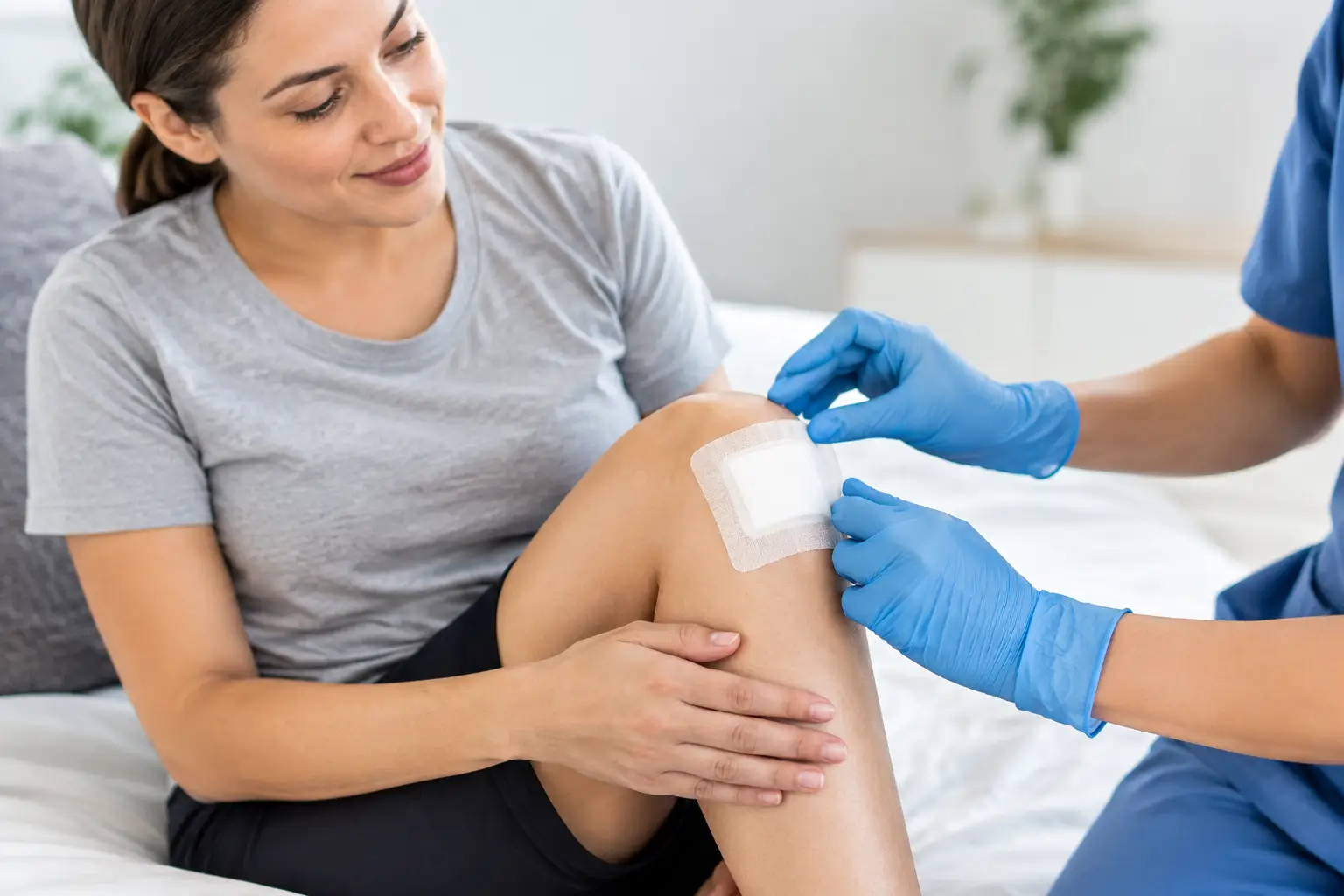
Keep the wound clean and dry as instructed, use the recommended dressing, and avoid stretching or friction at the site. Activities such as swimming, gym training and contact sports may need to wait until the wound has healed or sutures have been removed. Sun protection is particularly important while the scar matures, as ultraviolet exposure can make a new scar darker and more noticeable.
Contact your treating team if pain is increasing rather than settling, redness is spreading, there is discharge, the wound opens, or you develop a fever. These signs do not always indicate infection, but they should be assessed promptly.
A thoughtful approach leads to safer care
Mole removal is often a minor procedure, but the decision deserves careful attention. Whether the concern is diagnostic, practical or cosmetic, specialist assessment helps ensure the treatment method matches the lesion and that you understand the likely scar, recovery and pathology process. A calm consultation is the right place to ask questions, consider your options and choose care that protects both your health and your confidence.