A changing spot on the side of the nose can feel easy to dismiss until it begins to bleed after washing or forms a recurring crust. This skin cancer excision case example shows how a specialist-led assessment, careful surgery and pathology reporting work together to treat a suspicious lesion while giving proper attention to healing and scarring.
The case below is representative rather than a description of one individual patient. Every lesion, diagnosis and reconstruction plan is different. A consultation and, where appropriate, a biopsy are needed before treatment decisions can be made.
The case: a persistent lesion on the nose
A patient in their late sixties attended with a small pink, pearly lesion on the outer side of the nose. It had been present for more than a year and had gradually enlarged. It was not painful, but it would occasionally bleed when rubbed with a towel and had not settled despite moisturisers and over-the-counter treatments.
The patient had fair skin and a history of substantial sun exposure, but no previous skin cancer. On examination, the lesion had features that raised concern for a basal cell carcinoma, often called a BCC. BCCs are the most common form of skin cancer. They rarely spread elsewhere in the body, but can grow into nearby skin and tissue if left untreated. On the nose, even a small lesion needs thoughtful management because there is limited spare skin and the area is highly visible.
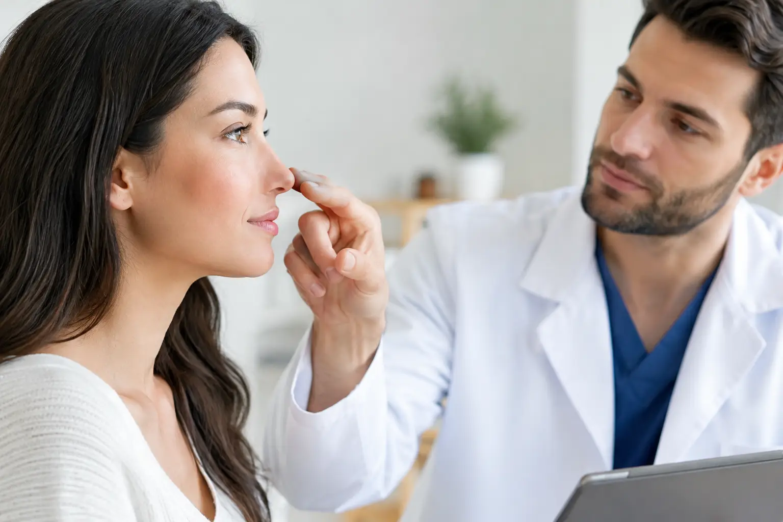
A specialist assessment considers more than whether a lesion looks suspicious. Its size, borders, location, how long it has changed, the patient’s medical history and any previous treatment all influence the safest approach. The clinician also examines the surrounding skin and may check regional lymph nodes when the type of lesion makes this relevant.
Deciding between biopsy and excision
In this example, the lesion was small enough and clinically suspicious enough for complete excision to be considered appropriate. In other circumstances, a small diagnostic biopsy may come first. This is particularly useful when the diagnosis is uncertain, when a lesion is large, or when the likely repair would be more involved and should be planned after the pathology result is known.
Excision means removing the visible lesion along with a measured rim of normal-looking skin around it. That rim is called a surgical margin. The appropriate margin depends on the suspected cancer type, its features and its location. A margin for a low-risk BCC will not necessarily be the same as one for a squamous cell carcinoma or melanoma.
This distinction matters. Skin cancer surgery is not simply about taking away a mark. The aim is to remove the lesion completely while preserving healthy tissue wherever safely possible. Occasionally, this balance means a staged procedure, a more specialised technique, or a repair that is more substantial than patients initially expect.
Preparing for skin cancer excision
The patient discussed the proposed procedure, alternatives, expected scar, possible need for further surgery if margins were involved, and practical aftercare. Medical conditions, medications and allergies were reviewed. In particular, blood-thinning medicines require individual advice. They should never be stopped without instruction from the clinician managing them.
For a small facial excision, treatment is often performed under local anaesthetic. The patient remains awake but should not feel pain in the area being treated. Some people feel pressure or movement during surgery, which is normal. If a lesion is larger, located in a complex area, or requires a more extensive reconstruction, a different setting or anaesthetic plan may be advised.
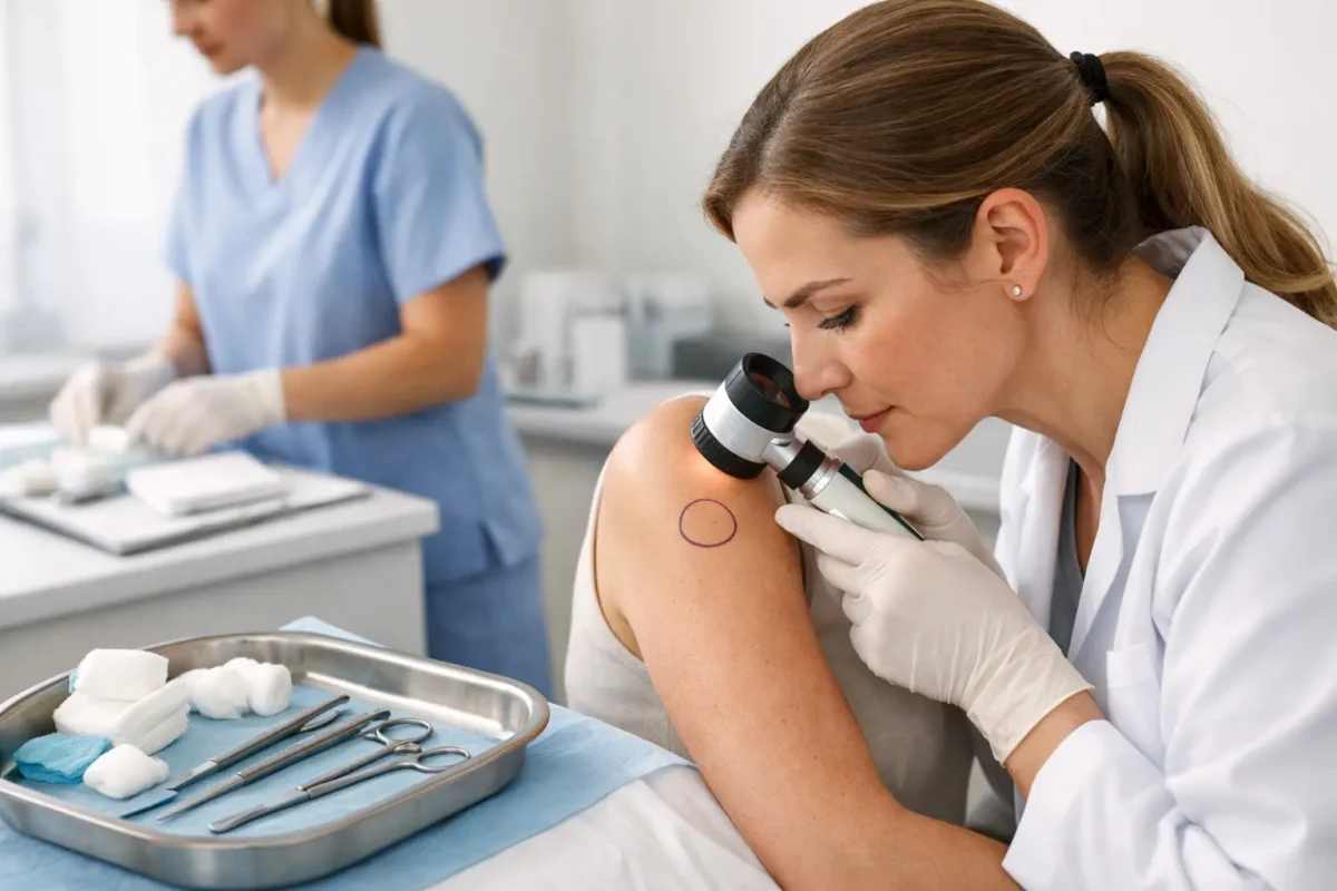
Photographs and measurements may be taken as part of the clinical record and surgical planning. This can feel formal, but it helps document the lesion accurately and supports clear follow-up care.
The procedure and reconstruction
After the local anaesthetic had taken effect, the lesion was removed with the planned margin and sent to the laboratory for histological examination. Histology is the microscopic assessment of tissue by a pathologist. It confirms the diagnosis and reports whether the cancer has been fully removed at the edges of the specimen.
The wound in this case could not be closed comfortably by simply bringing the edges together. Closing it under too much tension may distort the nostril or produce a less favourable scar. Instead, a small local skin flap was used. This involves carefully moving adjacent skin and tissue into the defect while maintaining its blood supply.
A local flap can sound alarming, but it is a well-established reconstructive technique. Its purpose is to restore contour and function, not just cover a gap. Depending on the site and size of the wound, alternatives may include direct closure, a skin graft, allowing the wound to heal naturally, or referral for a more complex reconstruction.
No reconstructive option is entirely without trade-offs. Direct closure may leave a longer line scar. A graft may differ slightly in colour or texture from nearby skin. A flap can create additional incision lines, although these are planned to sit as discreetly as possible. The most suitable approach depends on the individual anatomy and the amount of tissue removed.
What the pathology result showed
The pathology report confirmed a nodular basal cell carcinoma. Importantly, the report stated that the lesion had been completely excised with clear margins. This meant no further treatment was needed for that specific site at the time.
Clear margins are reassuring, but they do not mean a person can never develop another skin cancer. People who have had one BCC have a higher likelihood of developing further sun-related lesions elsewhere, so regular skin awareness and follow-up remain sensible.
Sometimes a pathology report shows that cancer cells extend to, or very close to, an edge of the specimen. The next step then depends on the cancer type, the precise wording of the report, the anatomical site and the patient’s circumstances. Further excision may be recommended. In selected situations, close observation or another treatment may be appropriate. This is why the pathology result, rather than appearance alone, guides the final management plan.
Recovery and wound care
The patient went home the same day with a dressing, written instructions and contact details for the surgical team. Mild discomfort, swelling and bruising are common after facial surgery. Simple pain relief is often sufficient, although each patient receives advice suited to their health needs.
Keeping the wound clean, protecting the dressing and avoiding activities that could pull on the repair are important in the early days. Smoking and nicotine products can impair wound healing and increase complications, particularly where a flap or graft has been used. Patients are usually advised to avoid strenuous exercise until the surgeon confirms it is safe to resume.
Sutures on the face are commonly removed within about a week, although timing varies with the repair and location. Scar redness and firmness can persist for several months. Scars generally mature gradually, not overnight. Once the wound is fully healed, sun protection is particularly important, as ultraviolet exposure can darken a healing scar and contributes to future skin damage.
Patients should contact their clinical team promptly if they develop increasing pain, spreading redness, pus-like discharge, fever, persistent bleeding, a dressing that becomes saturated, or a flap or graft that changes colour. Most healing concerns are manageable when reviewed early.
Follow-up is part of treatment
At review, the wound was healing appropriately, the nostril shape was preserved and the pathology result was discussed in plain terms. The patient was advised to monitor their skin for new or changing lesions and to seek review sooner rather than waiting for a routine appointment if something did not look right.
A useful habit is to look for a sore that does not heal, a spot that repeatedly bleeds or crusts, a mole changing in size, shape or colour, or a new lesion that looks noticeably different from others. These signs do not always mean cancer, but they deserve assessment.
Skin cancer excision is most effective when it is based on an accurate diagnosis, careful planning and appropriate follow-up. If you have noticed a persistent or changing lesion, arranging a specialist assessment can provide clarity and a treatment plan tailored to both your health and the area of skin involved.