A changing mole, a sore that does not heal, or a new lump can be worrying precisely because it is hard to judge from appearance alone. This guide to skin lesion biopsies explains how a small sample of skin can provide a clearer diagnosis, what your appointment may involve, and how a specialist plans treatment around both safety and the best possible scar.
Why a skin lesion biopsy may be recommended
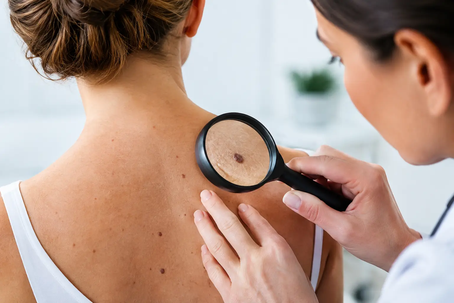
A skin lesion is an area of skin that looks or feels different from the surrounding skin. Many lesions are harmless, including cysts, skin tags, benign moles and age-related growths. However, some changes need closer assessment to rule out skin cancer or another condition that requires treatment.
Your doctor may recommend a biopsy when a lesion has changed in size, shape or colour; bleeds or crusts repeatedly; becomes painful or itchy; fails to heal; or has an appearance that cannot be diagnosed confidently during examination. A biopsy is not an assumption that a lesion is cancerous. It is a way of replacing uncertainty with an answer based on laboratory examination.
A specialist will consider the lesion’s appearance, location, size, duration and any history of previous skin cancers. Your personal and family history, sun exposure, medicines and tendency to form prominent scars may also influence the recommended approach.
What a biopsy can show
The sample is examined under a microscope by a pathologist. The report may identify a benign lesion, inflammation, infection, a precancerous change or a skin cancer. If cancer is found, the pathology helps guide the next step by describing the type of cancer and, where relevant, features that affect how much further treatment may be needed.
For some lesions, particularly small ones, the biopsy removes the whole area and no additional treatment is required. In other cases, a biopsy takes only part of the lesion because a diagnosis is needed before planning definitive excision or reconstruction. This distinction matters: removal of a lesion and a biopsy can occur in the same procedure, but they are not always the same thing.
Types of skin lesion biopsies
The right technique depends on the clinical question and where the lesion sits. The aim is to obtain an adequate sample while respecting function, healing and appearance.
Shave biopsy
A shave biopsy removes a superficial portion of a raised lesion using a fine blade. It can be suitable for selected benign-looking or superficial lesions. The wound often heals without stitches, although it may take several weeks to settle and can remain pink for a time.
Punch biopsy
A punch biopsy uses a small circular instrument to take a full-thickness core of skin. It is useful when the deeper layers of the skin need assessment, including for some rashes, inflammatory conditions and lesions where a representative sample is required. A small stitch may be used, depending on the size and location.
Incisional biopsy
An incisional biopsy removes a portion of a larger lesion. It may be chosen when taking the whole lesion immediately would create an unnecessarily large wound, or when the diagnosis will determine the extent of future surgery.
Excisional biopsy
An excisional biopsy removes the entire visible lesion, usually with a narrow margin of surrounding normal-looking skin. This is commonly considered for suspicious pigmented lesions when clinically appropriate. The specimen can then be assessed in full, while the wound is closed carefully with stitches.
What happens at the appointment
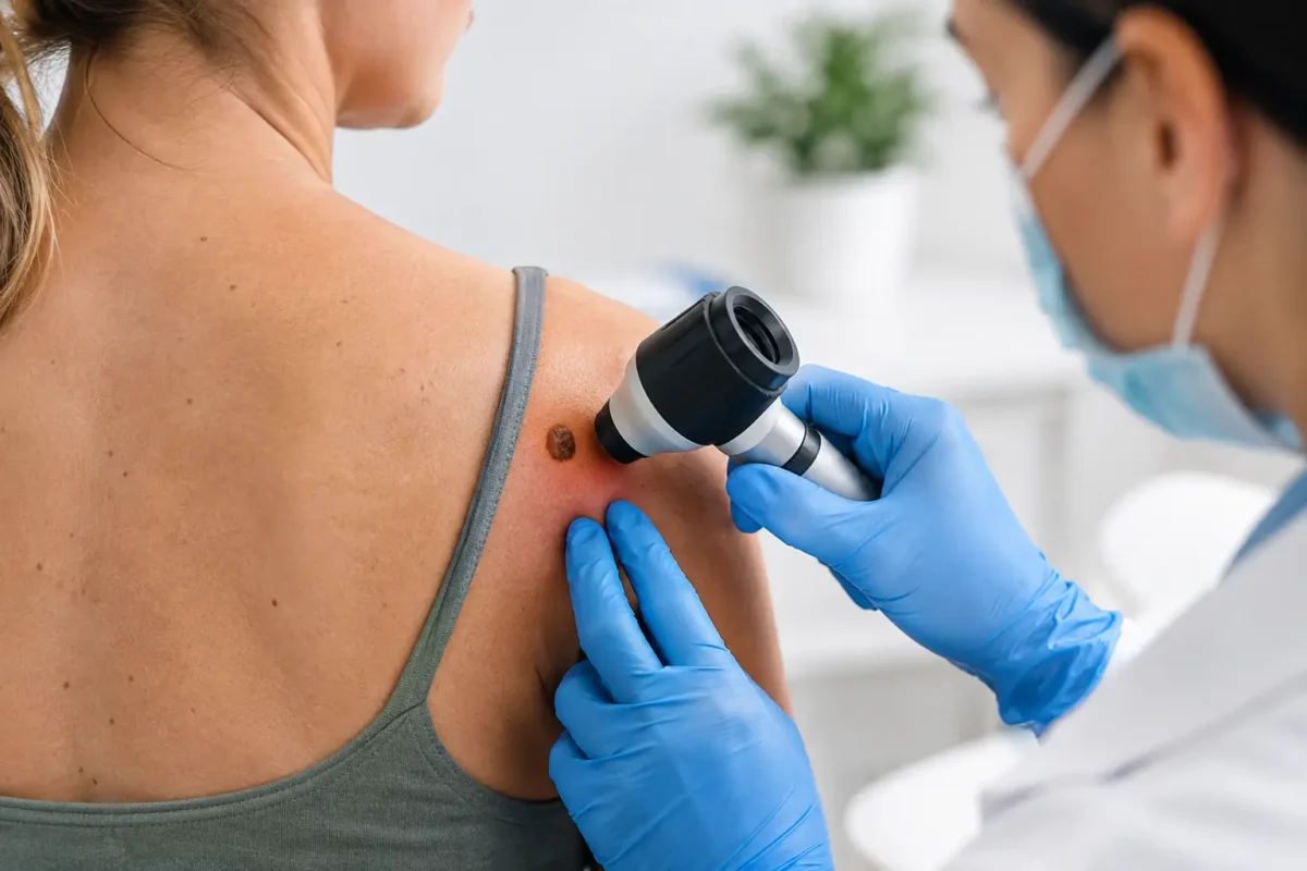
A consultation begins with a focused history and examination. The surgeon will discuss why a biopsy is recommended, the type of biopsy proposed, alternatives where appropriate, and what the resulting scar may look like. If the lesion is being removed from a cosmetically sensitive area, such as the face, ear, lip or eyelid region, closure planning is particularly important.
Most biopsies are performed under local anaesthetic. You will feel a brief sting as the anaesthetic is injected, after which the area should become numb. You may notice pressure or movement during the procedure, but you should not feel sharp pain. Let the clinical team know if you are uncomfortable at any stage.
The skin is cleaned, the sample is taken and the wound is dressed. Stitches may be dissolvable or may need to be removed at a follow-up appointment. Many minor procedures are completed within 20 to 45 minutes, although the total time in clinic can vary.
Before treatment, tell your surgeon about blood-thinning medication, allergies, diabetes, immune-suppressing medication, previous bleeding problems, pregnancy and any past difficulty with local anaesthetic. Do not stop prescribed medication unless the clinician managing your care advises you to do so.
Understanding results and next steps
Biopsy samples are sent for pathology assessment. Results are not usually available on the day of the procedure. The turnaround time varies according to laboratory workload and whether further specialist review of the sample is needed.
Your surgeon or clinical team will explain the result in clear terms and outline what it means for you. A benign result may need no further action beyond routine skin awareness. If margins are involved, if the diagnosis is uncertain, or if a skin cancer is identified, further excision or surveillance may be recommended.
When additional surgery is needed, it is planned according to the pathology result and the position of the lesion. This may involve removing a wider margin of skin or using a more considered closure technique to protect movement and minimise distortion of nearby features. Treatment should be individualised rather than based on a one-size-fits-all approach.
Healing after a biopsy
A small amount of tenderness, bruising, swelling or spotting on the dressing can be expected in the first few days. Simple pain relief is often sufficient, but follow the advice provided by your treating team. Keep the dressing clean and dry for the period advised, and avoid swimming, strenuous exercise or activities that pull on the wound until it is safe to resume them.
Scars mature slowly. Early redness and firmness are normal, and the final appearance may take many months to develop. Once the wound has fully healed, protecting the area from sun exposure can help reduce persistent colour change. Some sites, such as the chest, shoulders and upper back, are more prone to raised or widened scars. Your surgeon can discuss this before the procedure where relevant.
Contact your clinical team promptly if you develop increasing pain, spreading redness, warmth, pus-like discharge, ongoing bleeding, fever, or a wound that opens. These symptoms do not always indicate a serious problem, but they should be assessed rather than ignored.
Questions worth asking before your procedure
A good consultation should leave you clear about the reason for the biopsy and the practicalities of recovery. It can help to ask whether the whole lesion will be removed, whether stitches will be required, how you will receive the result, and what may happen if further treatment is recommended.
You may also wish to ask about the likely scar, any restrictions on work or exercise, and whether you need someone to accompany you home. Most local anaesthetic biopsies do not require this, but individual circumstances differ. Patients who feel informed before treatment are generally better prepared for the normal stages of healing and follow-up.
When not to wait
Arrange medical assessment for a mole or skin lesion that is changing, irregular in shape or colour, persistently bleeding, ulcerated, or new and growing. It is also sensible to seek review for a non-healing sore, particularly on sun-exposed skin. Early assessment does not mean the lesion is serious, but it allows the appropriate investigation to happen without unnecessary delay.
A biopsy is a short procedure, but its value can be significant: it provides the information needed to make a careful decision about treatment. If a lesion is concerning you, a specialist consultation can offer a clear plan, thoughtful surgical care and support through each stage of the process.