Categories
Plastic Surgery Skin Cancer

Skin Cancer: When a Changing Lesion Needs Review

https://plasticsurgerynz.org/skin-cancer-changing-lesion-review/
October 9, 2026 - Stanley Loo -A new mark that does not heal, or a mole that has begun to

A new mark that does not heal, or a mole that has begun to look different from the rest, deserves attention. Skin cancer is common, and most people will first notice it themselves, often while dressing, shaving, or applying moisturiser. Early assessment can make treatment simpler and can be particularly important for melanoma, the less common but more serious form of skin cancer.

Not every changing spot is cancerous. Ageing, sun exposure, irritation and harmless skin growths can all alter the appearance of the skin. The sensible response is not to assume the worst, but to arrange a timely clinical review when a lesion is new, changing, persistent or concerning.

What skin cancer can look like

Skin cancers do not all look alike. Some are pigmented and resemble an unusual mole. Others are pink, red, scaly, crusted, pearly or simply fail to heal. A lesion may be painless, so the absence of discomfort should not be used as reassurance on its own.

The three main forms are basal cell carcinoma, squamous cell carcinoma and melanoma. Basal cell carcinoma is the most common. It tends to grow slowly and rarely spreads elsewhere in the body, but it can cause significant local damage if left untreated, particularly around the nose, eyelids, ears and lips.

Squamous cell carcinoma may appear as a persistent rough or scaly patch, a thickened area, or a tender crusted growth. It can grow more quickly than basal cell carcinoma and, in a small proportion of cases, may spread beyond the skin. Prompt diagnosis and treatment are therefore advised.

Melanoma develops from pigment-producing cells. It can arise in an existing mole or appear as a new mark. It is sometimes brown or black, but may also be pink, red, blue-grey or skin-coloured. Melanoma is less common than other skin cancers, yet early diagnosis has a substantial effect on outcomes.

Skin cancer warning signs worth acting on

A useful way to assess a pigmented lesion is the ABCDE guide. Look for asymmetry, an irregular border, variation in colour, a diameter that is increasing, and evolution or change over time. Change is often the most meaningful feature: a mole that grows, darkens, becomes raised, bleeds or develops an unfamiliar appearance should be checked.

The “ugly duckling” sign is equally helpful. If one mole looks noticeably different from your usual pattern of freckles or moles, it is worth seeking advice even if it does not meet every ABCDE feature.

Non-pigmented lesions also require attention. Arrange an assessment if you notice a sore that has not healed after several weeks, recurring bleeding or crusting, a shiny bump, a steadily enlarging lump, or a rough red patch that persists despite simple skin care. A lesion that becomes painful, itchy or tender may need review, although many benign conditions can cause these symptoms too.

People sometimes delay because a mark is in a difficult-to-see place. The scalp, back, behind the ears, soles of the feet, nails and genital area should not be overlooked. Asking a partner, family member or hairdresser to mention a changing mark can be useful, but it should not replace a medical assessment.

Who has a higher risk?

Anyone can develop skin cancer, including people with darker skin tones. Risk rises with cumulative ultraviolet exposure, a history of severe sunburn, outdoor work or recreation, and use of sunbeds. Fair skin, light eyes, freckles, a large number of moles, a weakened immune system and a personal or family history of melanoma can also increase risk.

A previous skin cancer does not mean another will definitely occur, but it does justify regular skin awareness and follow-up tailored to the individual. Your clinician may advise a particular surveillance schedule depending on the type of cancer, its location and your wider medical history.

Sun protection remains worthwhile at every age. Shade, protective clothing, a broad-brimmed hat, sunglasses and broad-spectrum sunscreen all have a role. Sunscreen is helpful but should not be treated as a way to stay in intense sun for longer. Avoiding sunbeds is particularly important, as they expose the skin to concentrated ultraviolet radiation.

What happens at a specialist assessment?

A careful consultation starts with your history. You may be asked when you first noticed the lesion, whether it has changed, bled or become symptomatic, and whether you have had significant sun exposure or previous skin cancers. Your medications, allergies and relevant health conditions also matter when planning treatment.

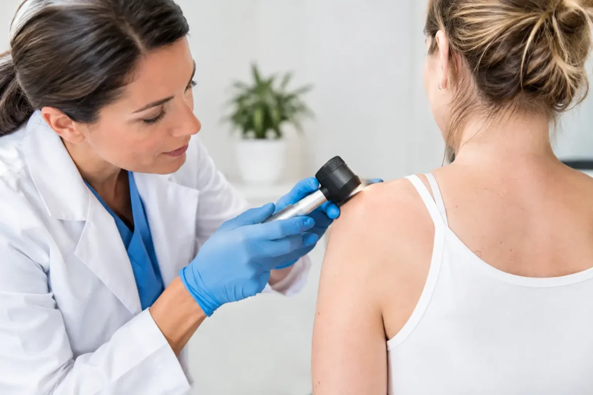
The skin lesion is examined closely, sometimes with a dermatoscope. This handheld instrument magnifies the lesion and uses light to show structures beneath the surface that cannot be seen easily with the naked eye. The surrounding skin may also be checked, particularly if you have numerous moles or sun-damaged areas.

Photographs can be useful for documentation and comparison, but they cannot reliably diagnose every lesion. A clinician may recommend observation for a clearly benign-looking mark, or removal and laboratory analysis where the diagnosis is uncertain or cancer is suspected. The right approach depends on the appearance, site and your individual risk factors.

Diagnosis and skin cancer excision

The definitive diagnosis usually comes from examining tissue under a microscope. In many cases, the entire lesion can be removed under local anaesthetic and sent to a laboratory for histology. This is known as an excision biopsy. For larger lesions or those in complex areas, a smaller biopsy may be taken first to guide further treatment.

Skin cancer excision involves removing the lesion with a measured margin of normal-looking skin around it. The margin is chosen according to the suspected diagnosis and site. This is why attempting to remove a suspicious lesion at home, or repeatedly freezing or treating it without a diagnosis, can complicate later assessment.

Once a lesion has been removed, the wound may be closed directly with stitches. If closing it directly would pull too tightly or distort an important feature, a skin graft or local flap may be considered. This is particularly relevant on the face, scalp, ears, hands and lower legs, where preserving function and achieving the best practical scar are both part of the surgical plan.

A plastic surgeon can be especially helpful where a cancer requires wider excision, where reconstruction is likely, or where the lesion lies near delicate structures such as the eyelid, nose, lip or ear. However, the priority is always complete and appropriate treatment of the cancer. Cosmetic considerations are important, but they should not compromise cancer clearance.

Understanding results and next steps

Laboratory results commonly confirm the type of lesion, whether it has been fully removed and whether further treatment is required. For some basal cell and squamous cell carcinomas, complete excision is the only treatment needed. If margins are involved, further surgery may be advised to reduce the chance of recurrence.

Melanoma results include details such as tumour thickness and ulceration, which help determine the next stage of care. A wider excision is often recommended after the initial diagnosis. Depending on the features of the melanoma, referral to a specialist multidisciplinary team and further investigations may be appropriate.

Waiting for results can feel unsettling. It is reasonable to ask when you can expect to hear from the clinic, who will discuss the findings with you, and what symptoms should prompt earlier contact. Clear communication is part of safe care, particularly when treatment plans may change after histology is available.

Recovery, scars and ongoing skin checks

Most minor excisions are day procedures performed under local anaesthetic. The area may feel sore or tight for a few days. You will usually receive guidance on wound care, bathing, activity, dressing changes and when stitches need to be removed. The advice varies according to the body site and type of repair.

A scar is an inevitable part of surgical removal, but its appearance usually improves over many months. Protecting the healing scar from sun exposure, avoiding unnecessary tension and following post-operative instructions can support the best possible healing. Scars can remain red, firm or raised for a time, especially in areas under movement or tension.

After treatment, regular self-checks help you become familiar with your skin. Choose a well-lit room and use a mirror for difficult areas. If you have a history of skin cancer, ask your treating clinician how often professional review is advisable. New or changing lesions should still be assessed, even if they arise years after successful treatment.

If a spot has changed or simply does not feel right, do not wait for it to become painful or dramatic. A calm, expert assessment can provide reassurance when a lesion is harmless and a clear treatment plan when it is not.

Categories
Plastic Surgery Skin Cancer

Advances in Skin Cancer Surgery for Patients

https://plasticsurgerynz.org/advances-in-skin-cancer-surgery/
September 25, 2026 - Stanley Loo -A skin cancer operation is not simply about removing a

A skin cancer operation is not simply about removing a visible mark. The surgeon must remove the cancer completely where possible, preserve healthy tissue, and plan a repair that heals reliably in an area that may be highly visible or functionally important. Advances in skin cancer surgery are helping surgeons achieve this balance with greater precision, while keeping the individual patient, tumour type and site firmly at the centre of decision-making.

For patients, the most meaningful progress is often not a single new technique. It is the combination of earlier assessment, accurate diagnosis, careful surgical margin planning, refined reconstruction and attentive follow-up. These steps can make a real difference to cancer clearance, comfort, scar quality and confidence during recovery.

Advances in skin cancer surgery: precision begins before the operation

The most appropriate treatment depends on what the lesion is, where it is located, how large it is, and whether it has features that suggest a higher risk of recurrence or spread. Basal cell carcinoma, squamous cell carcinoma and melanoma behave differently, so they should not be approached as if they were the same condition.

Modern assessment may include dermoscopy, a close examination of skin structures using magnification and specialised lighting. Photography can also be useful for documenting a lesion and monitoring change. Where cancer is suspected, a biopsy may be recommended before definitive treatment, particularly if the diagnosis will alter the extent or nature of surgery.

This preparation matters because surgical planning is more accurate when the diagnosis is clear. A small, low-risk basal cell carcinoma on the trunk may be managed very differently from a lesion near an eyelid, lip, ear or nose. Similarly, melanoma requires a planned excision margin based on its measured depth, rather than a one-size-fits-all approach.

Not every changing or unusual lesion is cancer, but new, bleeding, crusting, non-healing or changing marks deserve timely assessment. It is generally better to have a concerning lesion examined early than to wait for it to become larger and more difficult to treat.

More accurate tumour removal

One of the central advances is the increasingly tailored use of surgical margins. A margin is the rim of normal-looking skin removed around a tumour to reduce the chance that microscopic cancer cells are left behind. The right margin is not necessarily the widest possible margin. It should be sufficient for the cancer type and risk profile while respecting the surrounding anatomy.

For selected skin cancers in high-risk or cosmetically sensitive locations, margin-controlled surgery may be considered. In this approach, tissue is assessed in a way that helps confirm whether cancer remains at the edges before the final reconstruction is completed. Mohs micrographic surgery is a well-known example, often used for particular basal cell and squamous cell carcinomas, especially on the face or where previous treatment has failed.

However, Mohs surgery is not automatically the best treatment for every patient or every skin cancer. It requires specific facilities and expertise, and conventional excision remains highly effective for many lesions. The key question is not which technique sounds most advanced, but which option offers appropriate cancer control with the least unnecessary tissue loss.

For melanoma and some higher-risk squamous cell carcinomas, treatment may involve further staging, discussion with other specialists or additional procedures. A specialist surgeon can explain when surgery is appropriate as a stand-alone treatment and when wider multidisciplinary care is needed.

Pathology remains a vital part of the process

Even with careful planning, the final pathology report guides the next step. It confirms the diagnosis, describes features of the tumour and reports whether it has been fully removed. Occasionally, further excision is advised because cancer cells are found close to or at an edge of the specimen.

This does not necessarily mean anything was done incorrectly. Skin cancers can extend beyond what is visible on the surface, particularly in areas of sun-damaged skin or where a lesion has poorly defined borders. Discussing pathology clearly is an important part of safe, informed care.

Better reconstruction after cancer removal

Reconstructive planning has progressed substantially. The aim is not merely to close a wound, but to restore the area in a way that supports healing, function and appearance. This is particularly relevant for skin cancer surgery on the face, scalp, ears, hands and lower legs.

A straightforward wound may be closed directly with carefully positioned stitches. Where direct closure would pull too tightly or distort a nearby structure, a skin graft or local flap may be more suitable. A local flap uses neighbouring skin and tissue, moved while maintaining its blood supply, to cover the defect. It can provide a close match in colour and texture, which is often valuable on the face.

The reconstructive method is chosen after considering more than the size of the wound. Skin laxity, blood supply, medical history, smoking status, medications and individual healing tendencies all matter. A technically possible repair is not always the best repair if it carries an unacceptable risk of delayed healing or functional problems.

In some cases, a staged approach is sensible. This may allow confirmation of clear margins before a more complex reconstruction, or give tissues time to settle before a later refinement. Patients should feel able to ask why a particular method has been recommended and what scar, swelling and recovery are realistically likely to involve.

Local anaesthetic surgery and patient comfort

Many skin cancer excisions can be performed under local anaesthetic. This avoids a general anaesthetic and usually allows patients to return home on the same day. Modern local anaesthetic techniques, careful tissue handling and clear perioperative guidance can make the experience more comfortable than many people expect.

That said, local anaesthetic is not suitable for everyone or every procedure. A large reconstruction, a difficult anatomical site, significant anxiety or relevant health conditions may mean that another anaesthetic option is safer or more appropriate. These decisions should be individualised rather than based solely on convenience.

Good surgery also includes good communication. Knowing what will happen on the day, how to look after the dressing, when stitches will be removed and whom to contact with concerns can reduce uncertainty during recovery.

Scar care and follow-up have become more considered

No surgeon can promise a scarless result. Any operation that removes skin will leave a scar, and the final appearance develops over months rather than days. Nevertheless, thoughtful incision placement, precise closure and protection of delicate surrounding tissues can help achieve the best possible result.

Aftercare also plays a part. Depending on the operation, advice may include keeping the wound dry initially, avoiding strenuous activity or stretching around the repair, using sun protection once healed, and attending scheduled wound checks. If a scar becomes raised, tight, tender or bothersome, treatments such as silicone therapy, massage, steroid treatment or scar revision may occasionally be discussed.

Follow-up is also an opportunity to consider the wider picture. Having one skin cancer increases the importance of regular skin awareness and sun protection. This does not mean every new mark is alarming, but it does mean changes should not be ignored.

What patients should look for in a surgical consultation

A high-quality consultation should leave you with a clear understanding of the suspected diagnosis, the proposed treatment and the alternatives. It should also cover the possibility of incomplete excision, recurrence, bleeding, infection, wound separation, altered sensation and scarring. These risks vary considerably by procedure and body site.

It is reasonable to ask who will perform the procedure, whether tissue will be sent for pathology, how results will be communicated, and what follow-up is included. For complex facial repairs or lesions in functionally important areas, reconstructive expertise can be particularly valuable.

At Stanley Loo Plastic Surgery, the focus is on consultant-led assessment, carefully planned treatment and practical support before and after surgery. The purpose is not to rush patients towards a procedure, but to provide clear advice that reflects both the cancer concern and the person sitting in the consultation room.

If you have noticed a lesion that is changing, bleeding, crusting or failing to heal, arrange an assessment rather than trying to judge it from appearance alone. Early, considered care gives both you and your surgeon more options for treatment and reconstruction.

Categories
Plastic Surgery Skin Cancer

How to Prepare for Lesion Excision Safely

https://plasticsurgerynz.org/how-to-prepare-for-lesion-excision/
September 19, 2026 - Stanley Loo -A lesion excision is often a relatively small procedure,

A lesion excision is often a relatively small procedure, but good preparation can make the day calmer, safer and easier to recover from. If you are wondering how to prepare for lesion excision, the most useful starting point is to understand exactly what is being removed, why it is being removed and what your individual surgical plan involves.

Skin lesions include moles, cysts, changing pigmented marks, skin tags, lumps and areas of suspected skin cancer. Some can be removed under local anaesthetic in a treatment room; others require a more involved excision, reconstruction or sedation. The preparation is therefore not identical for every patient. Your surgeon’s instructions should always take priority over general advice.

Start with a thorough consultation

The consultation is where safe lesion excision begins. Your surgeon will examine the lesion, ask how long it has been present and whether it has changed, bled, become painful, itchy or crusted. They may also ask about previous skin cancers, sun exposure, family history and medicines.

Be ready to explain any change you have noticed. A clear timeline can be more helpful than patients realise. If the lesion has altered in size, shape, colour or sensation, say when this began and whether the change was gradual or sudden. Photographs taken over time can occasionally be useful, although they do not replace a clinical assessment.

Your surgeon will discuss whether excision is appropriate, the likely scar, the type of anaesthetic and whether the specimen will be sent for laboratory examination. Sending tissue for histology is common when a diagnosis needs confirmation or a suspicious lesion is being removed. Results may affect whether any further treatment or follow-up is recommended.

This is also the right time to be open about your priorities. For a lesion on the face, scalp, chest or another visible area, scar placement and wound healing matter. However, the safest removal of a concerning lesion may sometimes require a longer scar than patients expect. A specialist will explain the balance between complete excision, accurate diagnosis and the best achievable cosmetic outcome.

Share your health information before the procedure

A full medical history helps your surgical team plan appropriately. Mention allergies, past reactions to local anaesthetic, bleeding problems, heart or lung conditions, diabetes, immune suppression, pregnancy, and any history of difficult scarring or keloid scars.

Medication review is particularly important. Blood-thinning medicines, including prescribed anticoagulants and antiplatelet medication, can increase bruising or bleeding. Some supplements may also have an effect. Do not stop aspirin, warfarin, clopidogrel, apixaban or any other prescribed medicine without clear advice from the clinician who manages it and your surgeon. Stopping certain medicines suddenly can be more dangerous than proceeding with a little extra bleeding risk.

Bring an up-to-date list of all medicines, inhalers, creams, vitamins and herbal products. If you use a topical treatment near the lesion, ask whether you should continue it before surgery. It is also helpful to tell the team about previous surgery in the same area, radiotherapy or recurrent infections, as these can influence wound healing.

Follow instructions on food, drink and anaesthetic

Many simple lesion excisions are performed under local anaesthetic. For these procedures, patients can usually eat and drink normally beforehand unless they have been given different instructions. Having a light meal and staying well hydrated can help you feel more comfortable.

If sedation or a general anaesthetic is planned, fasting rules apply. You will receive specific guidance about when to stop eating and drinking. Follow this carefully, including rules about chewing gum, sweets and milk in tea or coffee. Fasting instructions exist to reduce the risk of complications during anaesthesia, so they should not be treated as optional.

Ask in advance which anaesthetic is planned if you are unsure. Local anaesthetic numbs the treatment area while you remain awake. You may feel pressure or movement, but you should not feel sharp pain. Let the team know immediately if you are uncomfortable during the procedure. Sedation can make you drowsy and relaxed, while general anaesthetic means you will be asleep. Both involve additional preparation and recovery arrangements.

Prepare your skin and clothing

Unless advised otherwise, have a shower or wash on the morning of your procedure and arrive with clean skin. Avoid applying make-up, fake tan, moisturiser, perfume or heavy creams close to the treatment area. These can interfere with skin preparation and surgical markings.

Wear loose, comfortable clothing that does not rub against the expected dressing or pull over the treated area. A button-front or zip-front top can be particularly practical after excision on the chest, shoulder, upper arm or back. For a facial lesion, choose something easy to change out of without catching the dressing.

Do not shave the area unless your surgeon specifically asks you to. Shaving can cause tiny cuts and irritation, which may increase infection risk. The clinical team can trim hair safely if needed.

Arrange the practical details before the day

A small amount of planning prevents unnecessary stress after surgery. If your excision is under local anaesthetic and you feel well, you may be able to travel home independently. Nevertheless, it can be reassuring to have someone available, particularly if the lesion is on an area that may feel sore or restrict movement afterwards.

If you are having sedation or a general anaesthetic, arrange a responsible adult to take you home and stay with you for the period advised by your surgical team. You should not drive, operate machinery, drink alcohol, make important decisions or sign legal documents until the effects of anaesthetic medication have worn off.

Consider your work and home commitments too. An office-based role may be manageable soon after a straightforward excision, but physically demanding work, gym sessions, swimming and contact sport may need to wait. Tension across a fresh wound can cause bleeding, wound separation or a wider scar. The amount of restriction depends on the lesion’s location, the size of the excision and whether reconstruction was required.

Before attending, make sure you have:

  • your medication list and relevant medical information;
  • comfortable clothing and any advised support garments;
  • transport arrangements if sedation or general anaesthetic is planned;
  • a clear plan for avoiding strenuous activity afterwards; and
  • a way to contact the practice if you have a concern once home.

Know what to expect immediately afterwards

Your surgeon or nurse will explain how to care for the wound, when the dressing can be removed or changed, and whether your stitches will dissolve or need removal. Written instructions are valuable, especially if you are anxious or have received sedation, as it is easy to forget details after a procedure.

Keep the dressing clean and dry for the timeframe you have been given. Do not assume that advice from a previous procedure applies to this one. Some wounds can be gently washed relatively early, while others need longer protection because of their location or closure method.

Mild discomfort, bruising, swelling and a small amount of spotting on the dressing can be expected. Simple pain relief may be recommended, but use only the medicines your team has advised. If you take blood-thinning medication, avoid adding over-the-counter products without checking first.

Contact your surgical team promptly if bleeding does not settle with firm pressure, pain becomes severe or increasingly uncomfortable, redness spreads, swelling rapidly worsens, the wound opens, or you develop a fever or feel unwell. These symptoms do not always mean there is a serious problem, but they should be assessed rather than ignored.

Allow the scar time to mature

A scar is an expected part of lesion excision. Early scars are often pink, firm or slightly raised before they gradually settle. This process can take many months. Protecting the healing area from sun exposure is particularly important, as ultraviolet light can darken a new scar and make it more noticeable.

Once the wound is fully healed, your surgeon may recommend moisturising, silicone-based scar care, massage or other measures depending on the site and your skin type. Do not start these treatments over an open wound unless advised to do so.

Thoughtful preparation gives you more than a smoother procedure day. It allows your surgeon to make decisions with the fullest possible picture of your health, supports careful healing and gives you confidence about when to seek help. If anything about your planned lesion excision is unclear, asking before the day of surgery is always the sensible next step.

Categories
Plastic Surgery Skin Cancer

Are Skin Biopsies Painful? What to Expect

https://plasticsurgerynz.org/are-skin-biopsies-painful/
September 15, 2026 - Stanley Loo -A biopsy is often recommended because a mole, patch, lump

A biopsy is often recommended because a mole, patch, lump or sore needs a closer look under the microscope. It is understandable to ask, are skin biopsies painful? For most people, the brief sting of the local anaesthetic is the most uncomfortable part. Once the area is numb, you should feel pressure or movement rather than sharp pain.

The prospect of any procedure can be unsettling, particularly if this is your first experience of minor surgery. Knowing what will happen, what sensations are normal and when to contact your surgical team can make the appointment feel far more manageable.

Are skin biopsies painful during the procedure?

A skin biopsy is usually carried out using local anaesthetic. This means you remain awake, but the small area of skin being treated is numbed. The injection can sting or burn for several seconds, much like other injections, before the anaesthetic takes effect. Your clinician will allow enough time for the skin to become fully numb and will check this before starting.

During the biopsy itself, you may notice pushing, pulling or light pressure. You should not feel cutting pain. If you do feel anything sharp or uncomfortable, say so straight away. More local anaesthetic can usually be given promptly. There is no benefit in trying to tolerate pain silently.

The type and location of the biopsy matter. A small punch biopsy, which takes a circular sample of skin, is often quick. A shave biopsy removes a raised or superficial area and may not require stitches. An excision biopsy removes the entire lesion with a narrow margin of surrounding skin, so it can involve a longer incision and sutures. Even with an excision, the procedure is generally comfortable once the local anaesthetic is working.

What can make a skin biopsy feel more uncomfortable?

Everyone experiences procedures differently. Anxiety can make normal sensations feel more intense, especially when the biopsy is on an area you cannot easily see, such as the back, scalp or face. If you have previously had difficulty becoming numb at the dentist or during another procedure, mention this at your consultation.

Some sites are more sensitive than others. The ears, lips, nose, fingertips, toes and genital area have dense nerve supplies and can be tender while the injection is being given. Inflamed, infected or very sore skin may also be more sensitive before it is numbed.

A clinician will take these factors into account. Careful injection technique, allowing the anaesthetic adequate time to work and checking your comfort throughout are all part of good procedural care. In some circumstances, a different setting or sedation may be discussed, although this is not needed for most routine skin biopsies.

What does a biopsy feel like afterwards?

As the anaesthetic wears off, usually over the next few hours, mild soreness or throbbing is common. Many patients describe it as a small cut, graze or bruise rather than significant pain. The degree of tenderness depends on the size of the sample, whether stitches were used and where the wound is located.

Areas that move or rub are often more noticeable. A biopsy on the shoulder may pull when you reach overhead; one on the lower leg can ache after a day on your feet; and a site beneath clothing or a bra strap may feel irritated. Facial wounds can be surprisingly comfortable because of the good blood supply, while wounds on the lower legs and feet may take longer to settle.

Simple pain relief is usually enough. Paracetamol is commonly suitable for post-procedure discomfort when taken according to the packet directions and your clinician’s advice. Ask before taking anti-inflammatory medication such as ibuprofen if you have been advised to avoid it, take blood-thinning medicines, have a history of stomach ulcers or have kidney problems. Do not stop prescribed anticoagulants unless the clinician managing your care has specifically instructed you to do so.

Caring for the biopsy site can reduce soreness

Your surgical team should give you individual wound-care instructions, as advice differs according to the biopsy method, dressing and body site. Keeping the dressing clean and dry for the recommended period helps protect the wound in its early stages. After that, gentle washing may be advised. Avoid scrubbing, soaking in baths, swimming or applying unapproved creams until you have been told it is safe.

If stitches have been used, it is sensible to limit stretching and strenuous exercise for the period advised. This can reduce bleeding, discomfort and the chance of the wound separating. Stitches on the face are often removed sooner than those on the trunk, limbs or scalp, but timing is individual.

A small amount of spotting on the dressing is not unusual. If bleeding occurs, apply firm, continuous pressure with clean gauze or a clean cloth for the time your clinician has recommended. Repeatedly lifting the dressing to check can interrupt clotting. If bleeding does not stop, seek clinical advice.

When should you contact your clinician?

Mild tenderness should gradually improve, not become increasingly severe after the first couple of days. Contact your clinic if you develop worsening pain, spreading redness, warmth, swelling, pus-like discharge, a fever, persistent bleeding or a wound that opens. These symptoms do not always mean there is an infection, but they should be assessed.

It is also worth getting in touch if a dressing causes a rash, you are unsure when to have stitches removed, or you have concerns about the way the wound is healing. A prompt question is always preferable to worrying at home.

Why a biopsy may still be the right next step

The discomfort of a biopsy is usually short-lived, while the information it provides can be highly valuable. A laboratory examination can distinguish between harmless changes, inflammatory conditions, infections and skin cancers that may need treatment. Although a lesion can sometimes look reassuring or concerning on examination, a biopsy can provide the diagnostic certainty needed to plan appropriate care.

For a suspicious mole or changing lesion, delaying assessment because of concern about pain is rarely helpful. Changes in size, shape, colour or sensation, bleeding without a clear reason, or a sore that does not heal should be assessed by an appropriately qualified clinician. The right approach depends on the lesion and your medical history, not just on how it appears in a photograph or online description.

Preparing for a calmer appointment

Before your procedure, tell the team about allergies, previous reactions to local anaesthetic, bleeding problems, diabetes, immune suppression and all medicines or supplements you take. Bring questions with you, particularly if the biopsy is on a visible area and you are concerned about scarring. Your clinician can explain the planned technique, likely scar direction, dressing requirements and when results are expected.

It can also help to wear comfortable clothing that gives easy access to the area and will not rub against the dressing afterwards. If you are prone to feeling faint during medical procedures, mention it before the appointment. This is common and allows the team to position and support you appropriately.

A skin biopsy is a small procedure, but it deserves the same clear communication and attentive follow-up as any other treatment. Feeling a short sting is normal; feeling unsupported or uncertain should not be. A careful discussion with your clinician can help you approach the appointment with realistic expectations and a clear plan for healing.