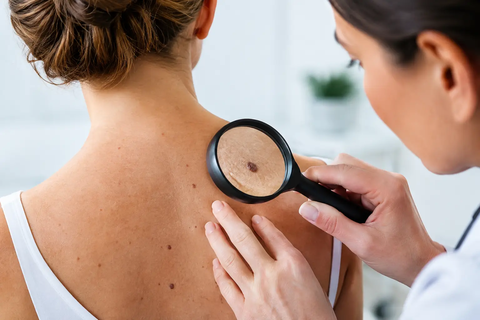
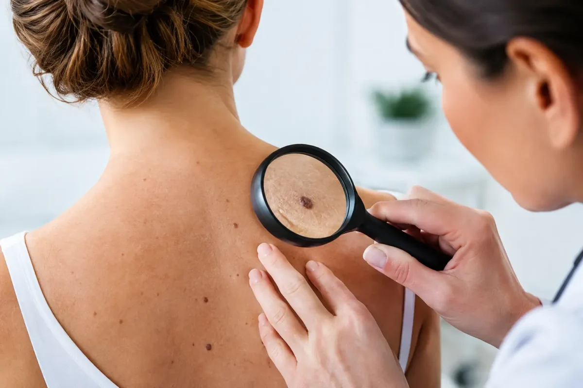
A new mark that does not heal, or a mole that has begun to look different from the rest, deserves attention. Skin cancer is common, and most people will first notice it themselves, often while dressing, shaving, or applying moisturiser. Early assessment can make treatment simpler and can be particularly important for melanoma, the less common but more serious form of skin cancer.
Not every changing spot is cancerous. Ageing, sun exposure, irritation and harmless skin growths can all alter the appearance of the skin. The sensible response is not to assume the worst, but to arrange a timely clinical review when a lesion is new, changing, persistent or concerning.
What skin cancer can look like
Skin cancers do not all look alike. Some are pigmented and resemble an unusual mole. Others are pink, red, scaly, crusted, pearly or simply fail to heal. A lesion may be painless, so the absence of discomfort should not be used as reassurance on its own.
The three main forms are basal cell carcinoma, squamous cell carcinoma and melanoma. Basal cell carcinoma is the most common. It tends to grow slowly and rarely spreads elsewhere in the body, but it can cause significant local damage if left untreated, particularly around the nose, eyelids, ears and lips.
Squamous cell carcinoma may appear as a persistent rough or scaly patch, a thickened area, or a tender crusted growth. It can grow more quickly than basal cell carcinoma and, in a small proportion of cases, may spread beyond the skin. Prompt diagnosis and treatment are therefore advised.
Melanoma develops from pigment-producing cells. It can arise in an existing mole or appear as a new mark. It is sometimes brown or black, but may also be pink, red, blue-grey or skin-coloured. Melanoma is less common than other skin cancers, yet early diagnosis has a substantial effect on outcomes.
Skin cancer warning signs worth acting on
A useful way to assess a pigmented lesion is the ABCDE guide. Look for asymmetry, an irregular border, variation in colour, a diameter that is increasing, and evolution or change over time. Change is often the most meaningful feature: a mole that grows, darkens, becomes raised, bleeds or develops an unfamiliar appearance should be checked.
The “ugly duckling” sign is equally helpful. If one mole looks noticeably different from your usual pattern of freckles or moles, it is worth seeking advice even if it does not meet every ABCDE feature.
Non-pigmented lesions also require attention. Arrange an assessment if you notice a sore that has not healed after several weeks, recurring bleeding or crusting, a shiny bump, a steadily enlarging lump, or a rough red patch that persists despite simple skin care. A lesion that becomes painful, itchy or tender may need review, although many benign conditions can cause these symptoms too.
People sometimes delay because a mark is in a difficult-to-see place. The scalp, back, behind the ears, soles of the feet, nails and genital area should not be overlooked. Asking a partner, family member or hairdresser to mention a changing mark can be useful, but it should not replace a medical assessment.
Who has a higher risk?
Anyone can develop skin cancer, including people with darker skin tones. Risk rises with cumulative ultraviolet exposure, a history of severe sunburn, outdoor work or recreation, and use of sunbeds. Fair skin, light eyes, freckles, a large number of moles, a weakened immune system and a personal or family history of melanoma can also increase risk.
A previous skin cancer does not mean another will definitely occur, but it does justify regular skin awareness and follow-up tailored to the individual. Your clinician may advise a particular surveillance schedule depending on the type of cancer, its location and your wider medical history.
Sun protection remains worthwhile at every age. Shade, protective clothing, a broad-brimmed hat, sunglasses and broad-spectrum sunscreen all have a role. Sunscreen is helpful but should not be treated as a way to stay in intense sun for longer. Avoiding sunbeds is particularly important, as they expose the skin to concentrated ultraviolet radiation.
What happens at a specialist assessment?
A careful consultation starts with your history. You may be asked when you first noticed the lesion, whether it has changed, bled or become symptomatic, and whether you have had significant sun exposure or previous skin cancers. Your medications, allergies and relevant health conditions also matter when planning treatment.
The skin lesion is examined closely, sometimes with a dermatoscope. This handheld instrument magnifies the lesion and uses light to show structures beneath the surface that cannot be seen easily with the naked eye. The surrounding skin may also be checked, particularly if you have numerous moles or sun-damaged areas.
Photographs can be useful for documentation and comparison, but they cannot reliably diagnose every lesion. A clinician may recommend observation for a clearly benign-looking mark, or removal and laboratory analysis where the diagnosis is uncertain or cancer is suspected. The right approach depends on the appearance, site and your individual risk factors.
Diagnosis and skin cancer excision
The definitive diagnosis usually comes from examining tissue under a microscope. In many cases, the entire lesion can be removed under local anaesthetic and sent to a laboratory for histology. This is known as an excision biopsy. For larger lesions or those in complex areas, a smaller biopsy may be taken first to guide further treatment.
Skin cancer excision involves removing the lesion with a measured margin of normal-looking skin around it. The margin is chosen according to the suspected diagnosis and site. This is why attempting to remove a suspicious lesion at home, or repeatedly freezing or treating it without a diagnosis, can complicate later assessment.
Once a lesion has been removed, the wound may be closed directly with stitches. If closing it directly would pull too tightly or distort an important feature, a skin graft or local flap may be considered. This is particularly relevant on the face, scalp, ears, hands and lower legs, where preserving function and achieving the best practical scar are both part of the surgical plan.
A plastic surgeon can be especially helpful where a cancer requires wider excision, where reconstruction is likely, or where the lesion lies near delicate structures such as the eyelid, nose, lip or ear. However, the priority is always complete and appropriate treatment of the cancer. Cosmetic considerations are important, but they should not compromise cancer clearance.
Understanding results and next steps
Laboratory results commonly confirm the type of lesion, whether it has been fully removed and whether further treatment is required. For some basal cell and squamous cell carcinomas, complete excision is the only treatment needed. If margins are involved, further surgery may be advised to reduce the chance of recurrence.
Melanoma results include details such as tumour thickness and ulceration, which help determine the next stage of care. A wider excision is often recommended after the initial diagnosis. Depending on the features of the melanoma, referral to a specialist multidisciplinary team and further investigations may be appropriate.
Waiting for results can feel unsettling. It is reasonable to ask when you can expect to hear from the clinic, who will discuss the findings with you, and what symptoms should prompt earlier contact. Clear communication is part of safe care, particularly when treatment plans may change after histology is available.
Recovery, scars and ongoing skin checks
Most minor excisions are day procedures performed under local anaesthetic. The area may feel sore or tight for a few days. You will usually receive guidance on wound care, bathing, activity, dressing changes and when stitches need to be removed. The advice varies according to the body site and type of repair.
A scar is an inevitable part of surgical removal, but its appearance usually improves over many months. Protecting the healing scar from sun exposure, avoiding unnecessary tension and following post-operative instructions can support the best possible healing. Scars can remain red, firm or raised for a time, especially in areas under movement or tension.
After treatment, regular self-checks help you become familiar with your skin. Choose a well-lit room and use a mirror for difficult areas. If you have a history of skin cancer, ask your treating clinician how often professional review is advisable. New or changing lesions should still be assessed, even if they arise years after successful treatment.
If a spot has changed or simply does not feel right, do not wait for it to become painful or dramatic. A calm, expert assessment can provide reassurance when a lesion is harmless and a clear treatment plan when it is not.