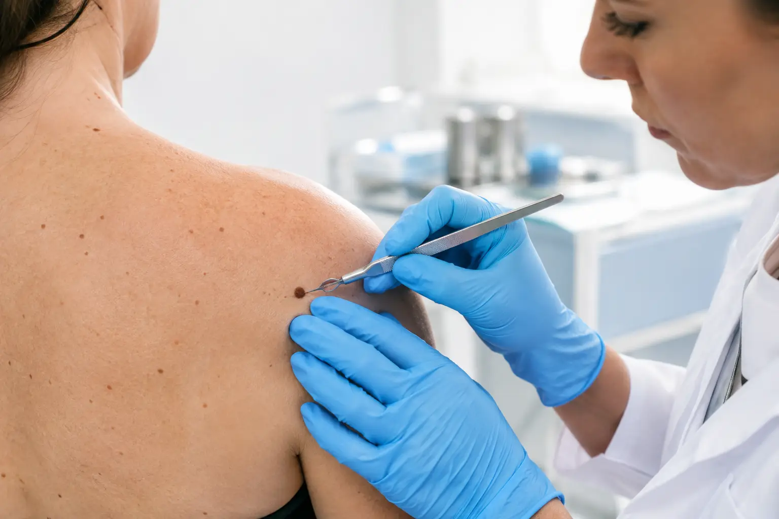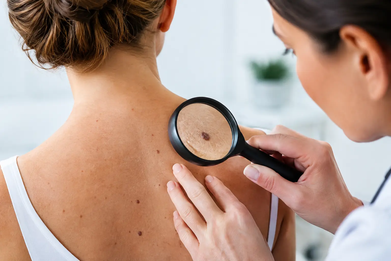
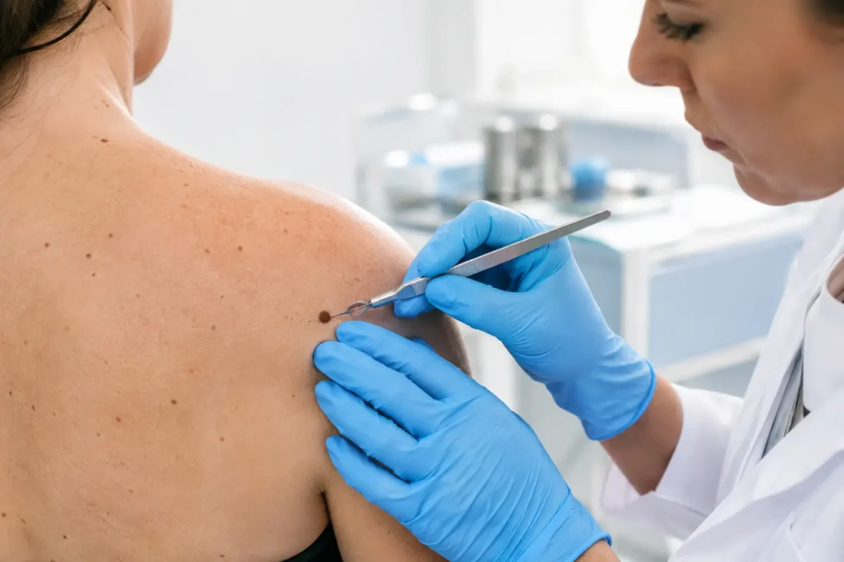
A changing mole, a persistent scaly patch or a lump beneath the skin can raise understandable questions. Surgical biopsy options allow your specialist to remove a small sample, or sometimes the whole lesion, so it can be examined under a microscope. The purpose is not simply to obtain an answer. It is to obtain the right answer while considering the lesion’s features, its location, the likely diagnosis and the best cosmetic and functional outcome.
A biopsy may be recommended when a lesion is new, changing, bleeding, painful, repeatedly irritated or clinically suspicious. Many biopsies confirm a benign condition. However, where skin cancer or another significant diagnosis is possible, a carefully planned procedure gives the pathologist the information needed to guide treatment.
When a surgical biopsy may be recommended
Your consultation begins with a detailed assessment of the area of concern. Your surgeon will ask how long it has been present, whether it has changed, and whether you have a personal or family history of skin cancer. They will examine the lesion’s size, shape, colour, border, texture and position, as well as the surrounding skin.
Some lesions have a clearly benign appearance and can be removed for symptoms or appearance without further investigation. Others need histological assessment because it is not possible to make a reliable diagnosis from appearance alone. A biopsy is also useful where a previous treatment has not resolved a lesion, or where an existing scar, lump or pigment change requires clarification.
The type of biopsy is chosen for a reason. Taking too little tissue can make interpretation difficult, while removing more tissue than necessary may create an avoidable scar. Your specialist will balance diagnostic accuracy with the practicalities of closure, healing and the importance of preserving normal function.
Surgical biopsy options and why they differ
Excision biopsy
An excision biopsy removes the entire visible lesion, usually with a small margin of normal-looking skin. It is commonly appropriate for small pigmented lesions, suspicious moles, cysts, skin cancers and lumps that can be removed safely in one procedure.
The tissue is sent to a pathology laboratory for analysis. If the lesion is benign and completely removed, no further treatment may be required. If cancerous or pre-cancerous changes are identified, the pathology report helps determine whether the margins are clear and whether further excision or other treatment is advisable.
Excision biopsy often provides the most complete specimen, which can be particularly valuable when melanoma is a concern. It does, however, leave a linear scar and may require stitches. The length and direction of the scar are planned carefully, particularly on the face, scalp, hands or near joints, where skin movement and tension affect healing.
Incision biopsy
An incision biopsy removes only part of a larger lesion. This may be the preferred approach when complete removal would be extensive, when the lesion is in a complex area, or when the diagnosis will determine the scope of later treatment.
For example, a large lesion may require a sample from the most representative area before planning definitive surgery. The specimen must be deep and appropriately located to give the pathologist useful information. An incision biopsy generally requires a small wound closure and leaves a smaller scar than immediate complete removal, but a second procedure may be needed once the result is available.
Punch biopsy
A punch biopsy uses a circular instrument to remove a small, full-thickness core of skin. It is often useful for rashes, inflammatory skin conditions, small lesions and selected pigmented or non-pigmented areas where a representative sample is needed.
Depending on its size and location, the small wound may heal naturally or be closed with one or two stitches. A punch biopsy is quick and usually performed under local anaesthetic, but it does not always remove the whole lesion. If results are inconclusive or show an area requiring wider treatment, further surgery may be recommended.
Shave or superficial biopsy
A shave biopsy removes a raised or superficial portion of a lesion. It can be suitable for certain benign growths and superficial skin abnormalities. Healing commonly occurs without stitches, although the site may take several weeks to settle and can remain pink or lighter or darker than the surrounding skin for a time.
This approach is not appropriate for every suspicious pigmented lesion. When the depth of a lesion is clinically important, particularly where melanoma is being considered, an excision biopsy is often more informative. Your surgeon will explain why a particular technique is or is not suitable in your circumstances.
What happens during the procedure?
Most minor surgical biopsies are carried out as an outpatient procedure using local anaesthetic. The skin is cleaned, the area is numbed and the biopsy is performed once you are comfortable. You may feel pressure or movement, but you should not feel sharp pain.
If stitches are required, these are selected and placed according to the body area and the tension on the wound. A dressing will be applied, and you will receive clear instructions on keeping the site clean and dry, managing discomfort and recognising signs that need review. For larger procedures, anxious patients or situations involving more complex reconstruction, different anaesthetic arrangements may be discussed during consultation.
The procedure itself is usually straightforward, but good planning matters. The placement of an incision can influence how a scar matures. Areas such as the eyelids, lips, nose, ears, hands and lower legs have particular anatomical considerations, and treatment should account for both diagnosis and long-term function.
Understanding pathology results
The tissue sample is examined by a pathologist, who provides a written report. Results are not always available immediately, as careful processing and microscopic assessment take time. Your surgical team will advise when to expect an update and how the result will be communicated.
Pathology may confirm a harmless lesion, such as a benign mole, cyst or seborrhoeic keratosis. It may identify a pre-cancerous change, basal cell carcinoma, squamous cell carcinoma, melanoma or another condition requiring follow-up. The report may also describe whether a lesion appears fully removed at its edges, known as the margins.
A clear margin can be reassuring, but it is only one part of the decision-making process. Further treatment depends on the diagnosis, the lesion’s depth and subtype, its location, your medical history and accepted clinical guidance. Occasionally, additional surgery is recommended even after an apparently complete removal to achieve an appropriate treatment margin or to manage a higher-risk diagnosis.
Recovery, scarring and aftercare
Minor tenderness, bruising, swelling or a small amount of spotting on the dressing can be normal in the early days. Paracetamol is often suitable for discomfort, unless your doctor advises otherwise. You should avoid strenuous activity, swimming and stretching the area until your surgeon confirms it is safe, particularly if the wound is under tension.
Scars develop gradually. They often look pink, firm or slightly raised initially, then tend to soften and fade over months. No surgeon can promise a scar-free result, but careful closure, wound care and protection from sunlight can support better scar maturation. Once the wound has healed, sun protection is especially important to reduce persistent pigment change.
Contact your surgical team promptly if you develop increasing redness, warmth, swelling, worsening pain, pus-like discharge, persistent bleeding, fever or a wound that opens. These symptoms do not always mean infection, but they should be assessed.
Questions to discuss at your consultation
A good consultation should leave you clear on why a biopsy is being advised and what will happen next. Ask whether the whole lesion can be removed, what scar to expect, whether stitches will be needed, how long healing may take and when pathology results are likely to be available. If there is a possibility of further treatment, it is reasonable to ask what that might involve.
At Stanley Loo’s practice, biopsy planning is approached with the same attention given to any surgical procedure: a clear diagnosis, careful technique, realistic discussion of risks and attentive follow-up. The right option is individual, and a specialist assessment can provide both clarity and reassurance when a skin lesion needs investigation.
If you have noticed a lesion that is changing or concerning, arranging an assessment sooner rather than later can help you move from uncertainty to a considered plan of care.